Main content
Fungi are a normal part of skin flora, but also a common cause of disease. Fungal skin infections can clinically be divided into superficial and deep mycoses.
1. Superficial mycoses
Superficial fungi live on the dead keratin layer of the skin. Symptoms are scaling, erythema and vesicles, usually more prominent at the borders of a lesion. Fungi can also elicit an allergic or ‘id reaction’, an itchy eruption of small blisters at a site distant from the fungal infection, often hands/fingers. Fungi on the skin can be divided into dermatophytes (threadlike, branching filaments) and non-dermatophytes, for example yeasts.
Dermatophytes causing superficial mycoses are members of the groups Trichophyton, Microsporum and Epidermophyton. Infections with these dermatophytes are called ‘tinea’ and are classified based upon site of infection.
Dermatophyte superficial infections
Tinea capitis
This infection is seen primarily in children and the elderly. Finding the source of infection (human or animal) and limiting the spread are important. Attention should be given to sharing combs, etc. Complaints depend on the fungus and the host response and vary from mild erythema and scaling to extensive infection with hair loss, redness, pustules and exudate, leading to destruction of follicles (scarring alopecia). Causes are M. canis, T. verrucosum (often from animals), T. tonsurans and T. schoenleinii. Culture helps in diagnosis, but should not delay treatment. Treatment is necessary to prevent permanent hair loss, and should be started immediately. Oral antifungal agents such as griseofulvin (treatment of choice), and an imidazole or terbinafin are given to ensure that the fungus in hair and follicles is eradicated.
Tinea barbae
This dermatomycosis is limited to postpubertal males. Severe inflammation with multiple pustules and sometimes abscesses and sinuses is seen. The causative organisms are often zoophilic, for example T. mentagrophytes. As in tinea capitis, oral treatment should be given.
Tinea corporis
Tinea on the trunk and extremities classically presents as ‘ringworm’, with redness and scaling at the periphery and clearing in the centre. T. rubrum is the major cause. Variants are tinea faciei (face) and tinea cruris (groin). The latter is often associated with tinea pedis (see below) and can be confused with candidiasis, eczema and erythrasma. Diagnosis is based on clinical findings and KOH examination.
Tinea manuum
Redness and scaling (‘eczema’) on only one hand should always raise the suspicion of a tinea. Eczema is usully located on both hands. A KOH examination can be used to differentiate. Treatment is usually topical.
Tinea pedis / athletes foot
This is one of the commonest infections. It is often diagnosed but in more than 50% the cause may be bacterial or a mixed infection. Its increasing prevalence is particularly related to an increase in wearing closed shoes. It can be divided into three categories:
- Interdigital, especially between the 4th and 5th toe. Symptoms include redness, peeling, maceration and fissuring. Bacterial colonization can lead to ‘maceration’ and odour. Maceration is often caused by bacteria alone, KOH can differentiate.
- Moccasin type, a more chronic variant with hyperkeratotic scaling of the sole, heel and sides of the foot, with sometimes fissures.
- Vesiculobullous, a highly inflammatory variant.
T. rubrum is the commonest cause. The vesiculobullous variant is often caused by T. mentagrophytes, a zoonotic pathogen, explaining the more inflammatory reaction. In general patients have few subjective complaints. Treatment is given to limit progression and to avoid the risk of secondary bacterial infection. Usually topical therapy is sufficient.
Tinea unguium / onychomycosis
Though a common infection in temperate climates, fungal infection of the nail is less prevalent in (sub) tropical countries. Yeasts and spores are the main causes. Except for the (finger) nail, the nail fold can also become infected, causing paronychia. Fungi cause only a minority of nail disease. KOH examination or culture should therefore be performed before (systemic) treatment is started.
Non-dermatophytes superficial infections
Malassezia furfur and Candida albicans are both part of the normal skin flora. They can cause several skin diseases due to overgrowth and environmental factors such as heat and humidity, and host factors such as individual susceptibility, immunodeficiency, antibiotic use, nutritional status, etc.
Malassezia furfur
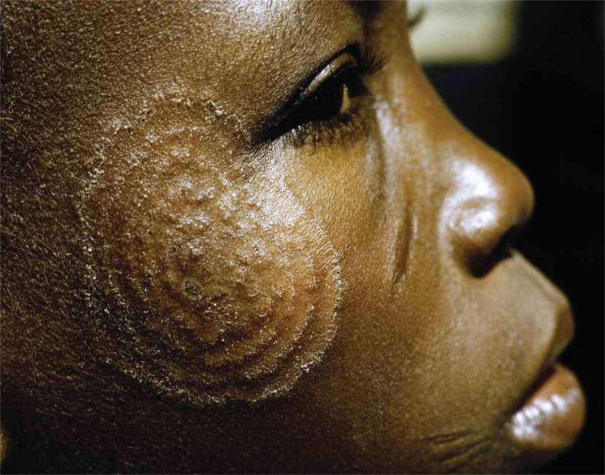
Pityriasis versicolor
This is a chronic, asymptomatic infection usually located on the trunk, neck and upper arms. Characteristically, it presents with hypopigmented (darker skin types) or hyperpigmented (lighter skin types), round to oval, sometimes confluenting, mildly scaly patches or plaques. To demonstrate the scaling, stretching the lesion is helpful. It is especially common in the tropics and subtropics and in HIV, sometimes occurring in up to 60% of the population. KOH examination shows the typical ‘spaghetti and meatballs’. Therapy can be given locally or systemically, depending on extent and cause. Relapses are common and must be differentiated from post inflammatory pigment changes. Scaling is a sign of active lesions.
M. furfur (pityrosporum) folliculitis
This is usually located on the upper half of the trunk. Itchy, monomorphic papules and pustules are seen. Microscopic evaluation with methylene blue or Gram stain confirms the diagnosis.Treatment is the same as for pityriasis versicolor.
Seborrhoic dermatitis
This is not a true fungal infection, but possibly an immunological reaction to M. furfur. Opinions about the role of M. furfur in seborrhoic dermatitis differ. It will not be discussed here.
Candida albicans
Candida intertrigo
Red patches with scaly, sometimes pustular borders. Satellite lesions are common. Predilection sites are the axillae, submammary, umbilicus, genital area, groin, peri-anal and the finger- and toe webs.
Differential diagnosis: intertriginous eczema, seborrhoic dermatitis and erythrasma. Bacterial superinfection is common.
Mucocutaneous candidiasis
Oral candidiasis: creamy, white flakes on a red, inflamed surface. The papillae may be atrophic or hypertrophic. Usually patients are immunosuppressed or use corticosteroid inhalers In candidiasis of the corners of the mouth, erythema and fissures can be seen. It can occur without underlying condition.
Vulvovaginal candidiasis
Usually itchy or burning erythema and ‘buttermilk-like’ vaginal discharge. Risk factors should be given attention to avoid relapses.
Onychomycosis and paronychia (see above).
2. Deep mycoses
Deep or subcutaneous mycoses are caused by a variety of fungi, that infect the skin including the subcutaneous tissue and in some instances underlying tissues and organs. See also ‘Tropical infectious ulcers’ and ‘Mycetoma’ in this special issue or the NVTG website.
Evaluation
Evaluation consists of scrapings of the ‘active’, red and scaly borders for KOH investigation. In onychomycosis the border of the affected nail can be used.
Culture is useful in the diagnosis of tinea capitis and onychomycosis. Treatment can be optimized, as some fungi, especially the zoonotic pathogens, are less sensitive to imidazoles.
Sometimes histopathology with PAS-D stain or Grocott silver is necessary. This is especially useful in deep mycoses.
One always has to bear in mind that due to, for example, inadequate treatment or host factors like immunosuppression, mycoses can lose their typical features. This is called ‘tinea incognito’. KOH examination or histopathology can be used for diagnosis.
Treatment
Most superficial dermatomycosis can be treated topically with an imidazole cream or Whitfield’s ointment. Usually the therapy should be given for at least 4 weeks or even better until one week after redness and scaling has disappeared.
Oral candidiasis can be treated with nystatin suspension, vaginal candidiasis with imidazole pessaries and cream.
If hair or nails are involved, or if the superficial infection is too extensive, systemic treatment should be given, for example griseofulvin (not for Candida!), terbinafin or one of the imidazoles. Treatment should only be stopped after complete clearance. Re-evaluating the patient at the end of treatment is therefore important.
Deep mycoses are very difficult to treat and usually require a combination of surgical debridement and systemic imidazoles.