Main content
The general health attendant in resource-poor countries is increasingly confronted with patients complaining of ants running under their skin, of aches and pains in face or limbs, or about loss of sensation in patches or in hand or foot. The area feels numb, dead, or has ‘pins and needles’.
The patient may present with hypopigmented or erythematous macules or papules and nodules or plaques which are skin coloured or slightly red. The health worker may think of a diagnosis, but often has to guess and the patient may become worse. Usually leprosy is not suspected; since it is often not included in the curriculum and there is no supervision of a leprosy service, because this has been dismantled, as a result of the claim of WHO that leprosy since 2005 has been eliminated as a worldwide Public Health problem. The alternatives, dermatologists and neurologists, are hardly trained in leprosy, are often not interested and not available. It may have been better in the early 1990s, when knowledge was still available to make a clinical diagnosis and to detect and treat complications in time.
This paper is meant to make the reader aware that leprosy is still present, to help diagnose and treat leprosy, and to recognize and treat complications.
Diagnosis of leprosy
Most important in the diagnosis is awareness. Clinically, leprosy is diagnosed when the patient shows two out of three signs. For endemic countries one is considered enough.
The three signs are:
- Loss of sensation in a skin lesion
- Enlarged nerve
- Positive skin smears
Loss of sensation is tested with a wisp of cotton wool; not with a ballpoint. The area in the lesion is tested by touching. With closed eyes the patient points to where he is touched. To make sure, the area outside the lesion is tested as well.
Enlarged nerves can be cutaneous nerves, subcutaneous nerves in the vicinity of skin patches or nerve trunks. Palpate at least the posterior auricular nerves, the ulnar nerves, the radiocutaneus nerves, the median nerves, the lateral popliteal nerves and the tibial posterior nerves. You can expand to every palpable nerve. Feel for thickness, consistence and tenderness.
Smears are taken to detect acid fast bacilli from the ears and other colder areas and from the rim of the lesion in paucibacillary (PB) patients and central in the lesion in multibacillary (MB) patients. The smear is taken with a surgical blade, while squeezing the skin, to numb and to diminish the bleeding; only tissue fluid is required. The cut is smeared with the blade 90 degrees on the cut direction, allowed to dry and stained for acid fast bacilli. The number of bacilli is read and graded along a logarithmic scale (BI: bacillary index) and the percentage of solid bacteria, these are live (viable) bacilli, is estimated (MI: morphological index).
Laboratory investigations are of limited help in the diagnosis of leprosy. However, the skin smear certainly is. Of some help is the titre against phenolic glycolipid 1 (PGL-1), a cell wall species specific glycolipid. However, this can be positive in contacts, and negative in paucibacillary leprosy. It is used by some to replace the smear. It helps to classify leprosy in PB and MB, and it can be used to follow the effect of treatment in MB patients and to detect relapses. Lymphocyte transformation tests against different antigenic determinants have been a disappointment up to now. PCR and NASBA are often negative in paucibacillary leprosy. They can be useful in the follow-up and the detection of relapses. Biopsy for histopathology can be very helpful, as can immunopathology, but the latter is only experimental. A problem can be that even within lesions, the histopathology of one spot may differ from the other. However it may give an idea of which immunological process goes on.
Infection and classification
Leprosy is highly infectious, but the attack rate is low. The major reason of this low attack rate is that most people genetically are unable to supply the mycobacteria in their cells with what they need to survive, because they lack the genes the bacterium needs.
In order to predict complications and to stratify along cell mediated immunity (CMI), the Ridley-Jopling scale is important, with on one side of the spectrum TT (polar tuberculoid) leprosy with a single well described lesion or an enlarged nerve, with no bacilli detectible and a high CMI against M. leprae antigenic determinants. On the other side there is LL (polar lepromatous ) leprosy with nodules and or plaques, symmetrical enlarged nerves or even only an infiltrated skin all over (Lepra bonita), with an absence of CMI against M.leprae antigenic determinants and many bacilli. In between is the borderline group, which comprises the majority of the patients. Borderline tuberculoid (BT) with predominantly tuberculoid features or borderline lepromatous (BL) with predominantly lepromatous features. Between those two is an unstable group of mid-borderline (BB) patients with typical, punched out or dome shaped lesions.
For fieldwork purposes, to make it simpler, one just counts the number of lesions: 5 or less is paucibacillary leprosy, more than 5 multibacillary leprosy.
Treatment
Multi Drug Therapy (MDT): Paucibacillary leprosy: 600 mg rifampicin once monthly under supervision and daily 100 mg dapsone for 6 monthly doses in 9 months’ time. The dose is for a 60 kg patient.
Multi bacillary leprosy: 600 mg rifampicin and 300 mg Lampren (clofazimine) once monthly under supervision and 100 mg dapsone and 50 mg Lampren daily. Twelve monthly doses should be given within 18 months for low BI patients, 24 monthly doses in 36 months for patients with a BI of 4 or more. The doses are for 60 kg patients.
Be careful with dapsone in Nordic Caucasians who easily develop haemolysis.
The treatment is effective, hardly any relapses are seen.
Reactions
Reactions belong to the normal course of a leprosy infection. However, treatment can prevent or precipitate them. There are 3 types of reactions; Type I leprosy reaction, also called Reversal Reaction (RR), type II leprosy reaction, also called Erythema Nodosum Leprosum (ENL) and Lucio’s phenomenon.
Type I reaction is a CMI reaction, a type IV Gell and Coombs reaction against M. leprae antigenic determinants. Clinically, there is increased inflammation of lesions, which become visible and erythematous, are raised or enlarged; they may even ulcerate. Nerves may be inflamed, enlarged and tender, with diminishing strength, sweating and sensitivity. There may be acro-edema.
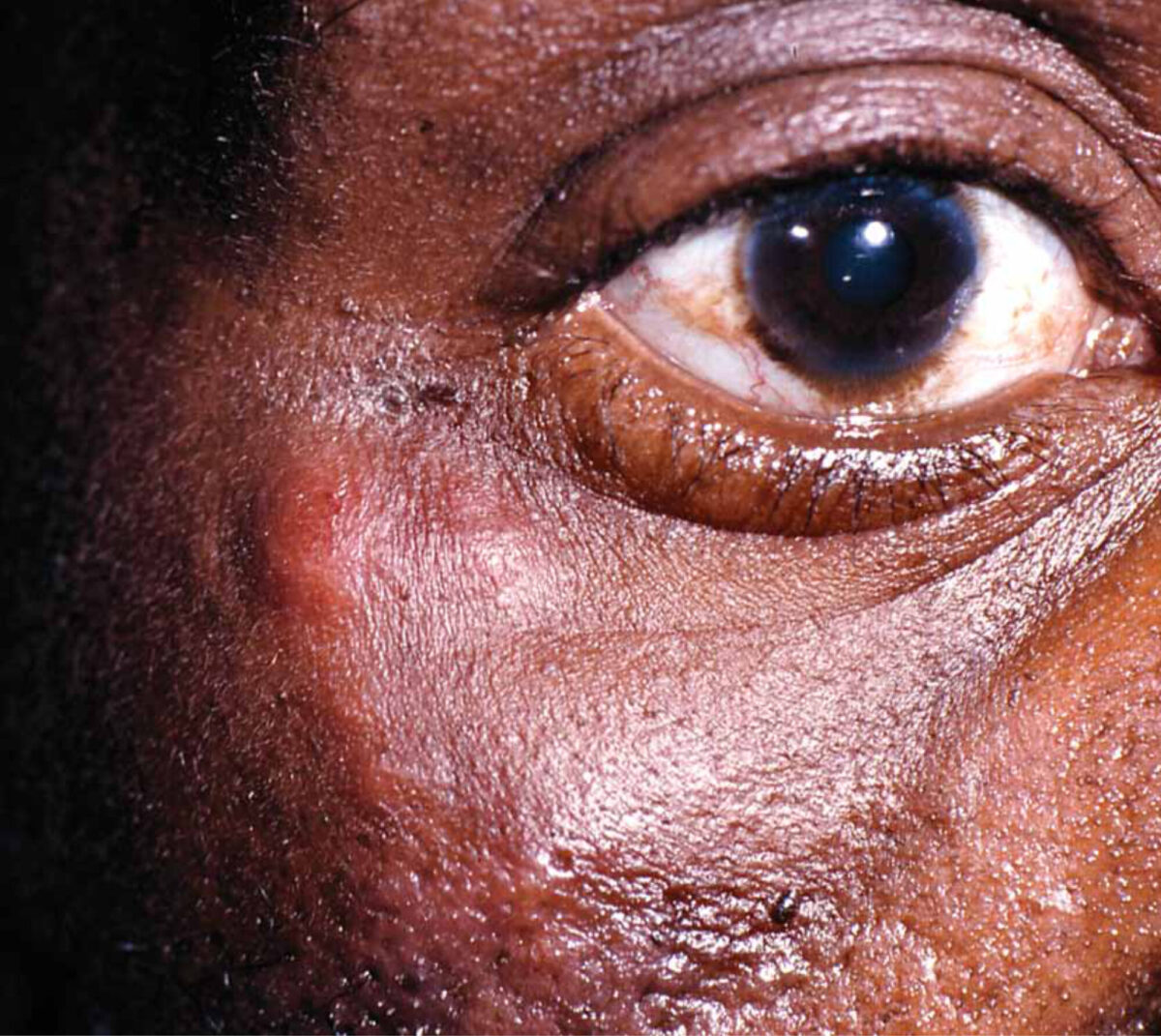
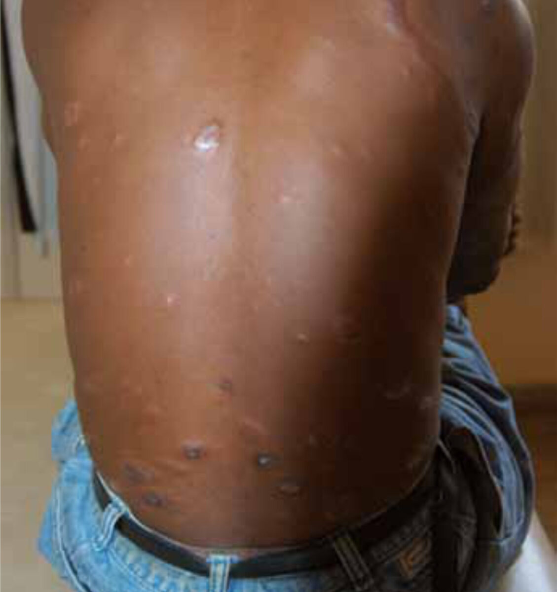
Type II leprosy reaction is an antigen-antibody immune complex reaction in the tissues, particularly in skin and nerve. The skin shows the characteristic red, painful, tender nodules. However, all organs can be involved and all tissues can be inflamed. There may be fever and leucocytosis.
The treatment of Type I reaction is primarily corticosteroids, 30-40 mg starting dose, tapering down, guided by, for instance, graded sensory testing, in 6-12 months, in which the dose needs to be 20 mg at least to be effective. In some cases dapsone helps to prevent a reaction.
Type II reaction treatment is difficult. The reaction is episodic, 95% of ENL episodes last less than one month. Mild reactions can be treated with NSAIDs; arthritis with antimalarials, but severe reactions need high dose steroids (60-120 mg) for a short period, diminishing to zero in a month or less. A new attack should be treated the same way. Lampren may prevent a Type II reaction or can be used as treatment. Thalidomide as treatment is superior above anything and can be used as prevention.
When nerves continue to deteriorate despite proper medical treatment, a nerve release operation needs to be considered. This can also be done for nerves without a reaction but which remain tender after treatment.
The Lucio phenomenon presents as an infarction of the skin, due to bacilli blocking the venous return. This is only seen in untreated leprosy. The treatment is MDT.
The results of nerve damage, loss of sensation and muscle strength are the sequelae or the stigmata of leprosy. These should be countered with supplying special, padded tools, utensils and shoes. Sometimes in order to increase grip or to improve foot movement, a tendon transfer may be considered, but always with an experienced physiotherapist present.
References
- Leprosy, A practical guide. Editors: Enrico Nunzi and Cesare