


Main content
Mycetoma (Madura foot) is a thoroughly neglected condition. It occurs worldwide, but Sudan seems to be the homeland. It affects mainly poor people in remote areas which may explain why it does not appear on any list of neglected tropical diseases and is not considered a priority by health authorities. However, it causes considerable chronic morbidity and often it leads to amputation of limbs after years of suffering; in severe cases, there is also mortality. As, in principle, it can be treated by simple medical treatment with limited surgery if needed, it deserves far more attention.
Mycetoma is a chronic infection of subcutaneous tissues that leads to disfigurement and disability in many parts of the world. It is caused by fungi or bacteria, hence it is called eumycetoma or actinomycetoma, respectively.
Eumycetoma is mostly caused by Madurella mycetomatis, while Nocardia spp. and Streptomyces somaliensis are the most common pathogens in Actinomycetoma.
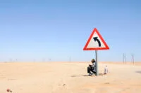
The disease is prevalent in the so-called mycetoma belt which stretches between latitudes of 15° South and 30° North, including Sudan, Somalia, India, Mexico, Venezuela and Argentina. These areas are relatively dry with a short rainy season of 4-6 months duration. In the dry season temperatures are 45-60°C during the day, 15-18°C at night with a relative humidity of 12-18%.
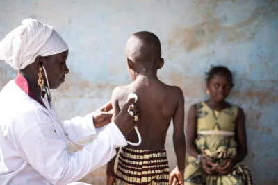
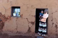
All ages can be affected but in endemic areas most patients present between 20-40 years of age thus affecting the most productive age group. Most patients are farmers or herdsmen; in a recent study in Sudan, however, 30% of patients were students and schoolchildren. The true incidence of mycetoma is not known; in many endemic areas mycetoma occurs in remote areas and many patients lack education or financial means to report to a hospital for treatment; others may fear amputation of the affected limb.
Worldwide most cases occur in Sudan; these are mainly eumycetoma and in 70% of these M. mycetomatis is the causative agent. The Mycetoma Research Centre in Khartoum has more than 6000 patients under treatment.
In a recent field study in a village in the endemic area in central Sudan the prevalence was 8.3/1000 inhabitants; eumycetoma was the third commonest health problem after malaria and schistosomiasis. The disease presentation included the whole spectrum from small nodules to extensive lesions and amputated limbs. Few patients had access to medical treatment; surgery was the rule.
The exact source of the infection is unknown; it is thought to be transmitted through a thorn-prick in areas where people tend to walk barefoot; the disease may, however, also affect other parts of the body. To date, the fungus has not been isolated from the soil or other sources. In the endemic villages most cases seemed to occur in the most densely populated part of the village where people live in the same compound as their animals (sheep, goats, dogs, chicken, donkeys) and the ground is covered with animal dung.
Clinical presentation
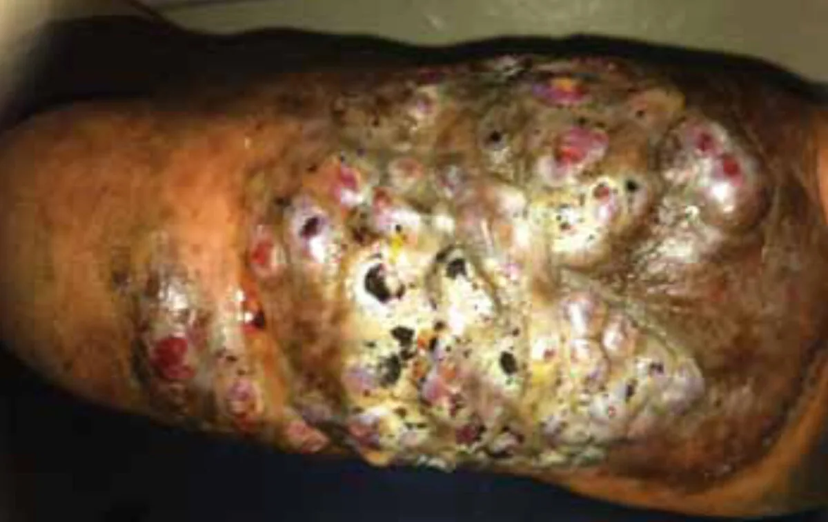
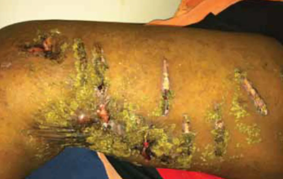
The typical clinical triad consists of a painless subcutaneous mass, sinus formation and purulent discharge that contains fungal grains. The lesion is usually on the lower limb, involving the foot. From there local spread occurs to lymph nodes and bone; haematogenous metastatic spread may also occur to other parts of the body. Mycetoma may also occur on the hands, and to a lesser extent on the chest, knee, head and neck and perineum. Pain is not a feature unless there is bone invasion or secondary bacterial infection; in one study the latter occurred in 65% of eumycetoma patients. The most common bacteria causing superinfection are S. aureus (56%), S. pyogenes (34%) and P. mirabilis (10%).
Diagnosis
The causative organism may be suspected from the colour of the grains; for example the grains are black in M. mycetomatis, red in Actinomadura pelletieri, white in Acremonium and Fusarium spp. and yellow in Streptomyces somaliensis and some Nocardia spp.
Definite confirmation is essential as this affects treatment; the organism may be identified by fine needle aspirate and examined by cytology, culture and PCR.
Imaging is useful to assess the extent of the lesion; this is done by X-ray, ultrasound, or MRI scan; clearly these techniques are only available to a certain extent in (referral) hospitals.
There is no rapid and reliable diagnostic test for use under field conditions and diagnosis will be mainly clinical; this may be problematic, particularly in early lesions.
Treatment
Treatment is basically with antifungals (eumycetoma) or antimicrobials (actinomycetoma), and surgery as appropriate. Actinomycetoma is treated with antibacterial agents such as cotrimoxazole; in extensive lesions aminoglycosides are added such as amikacin; the duration may be several months. While medical treatment in actinomycosis is usually satisfactory, in eumycetoma this is not the case. Current antifungals used include ketoconazole and itraconazole; however, these need to be given for 12 months. After that the lesion will have reduced in size and will be more accessible for non-mutilating surgery. However, often the fungus may still be cultured from the lesion after treatment and the recurrence rate is high. In addition, the cost of treatment is high and many patients drop out from follow-up early during treatment. All too often over time this leads to amputation of the limb after years of suffering and disability. In remote areas surgery is the primary treatment.
Newer antifungal agents such as voriconazole, posaconazole and isavuconazole are promising; in vitro studies show excellent activity. In contrast to ketoconazole and itraconazole, activity against voriconazole is not inhibited in its action by melanin produced by the fungus, suggesting better clinical activity. In vivo experience is so far limited to case reports but shows favourable results. Echinocandins are not effective against M. mycetomatis.
Issues in mycetoma control include
- Assessment of disease burden
- Advocacy for acknowledgement as a neglected tropical condition by WHO
- Health education
- Rapid and reliable diagnosis
- Affordable and safe medical treatment followed by surgery if necessary; explore concomitant antibacterial treatment in eumycetoma
- Research as to the source of the organism and mode of transmission
References
- Ahmed AOA, Abugroun EAM, Fahal AH, Zijlstra EE, van Belkum A, Verbrugh HA. Unexpected High Prevalence of Secondary Bacterial Infection in Patients with Mycetoma. J Clin Microbiol 1998;36:850-1.
- Ahmed AOA, van Leeuwen W, Fahal A, van de Sande W, Verbrugh H, van Belkum A. Mycetoma caused by Madurella mycetomatis: a neglected infectious burden. Lancet Infectious Diseases 2004;4:566-74.
- Van de Sande WW, de Kat J, Coppens J, Ahmed AO, Fahal A, Verbrugh H, van Belkum A. Melanin biosynthesis in Madurella mycetomatis and its effect on susceptibility to itraconazole and ketoconazole. Microbes Infect. 2007;9:1114-23.
- Van Belkum A, Fahal AH, van de Sande WW.In vitro susceptibility of Madurella mycetomatis to posaconazole and terbinafine. Antimicrob Agents Chemother 2011;55:1771-3.
- Van de Sande WW, Fahal AH, Bakker-Woudenberg IA,