Main content
Setting
This case is set in the Lion Heart Medical Centre in Yele, a rural hospital in the middle of Sierra Leone. The hospital is run by local staff and three tropical doctors trained in the Netherlands. They have access to a laboratory, ultrasound, and an X-ray machine (which is currently out of order). Furthermore, the hospital has an operating theatre.
Case
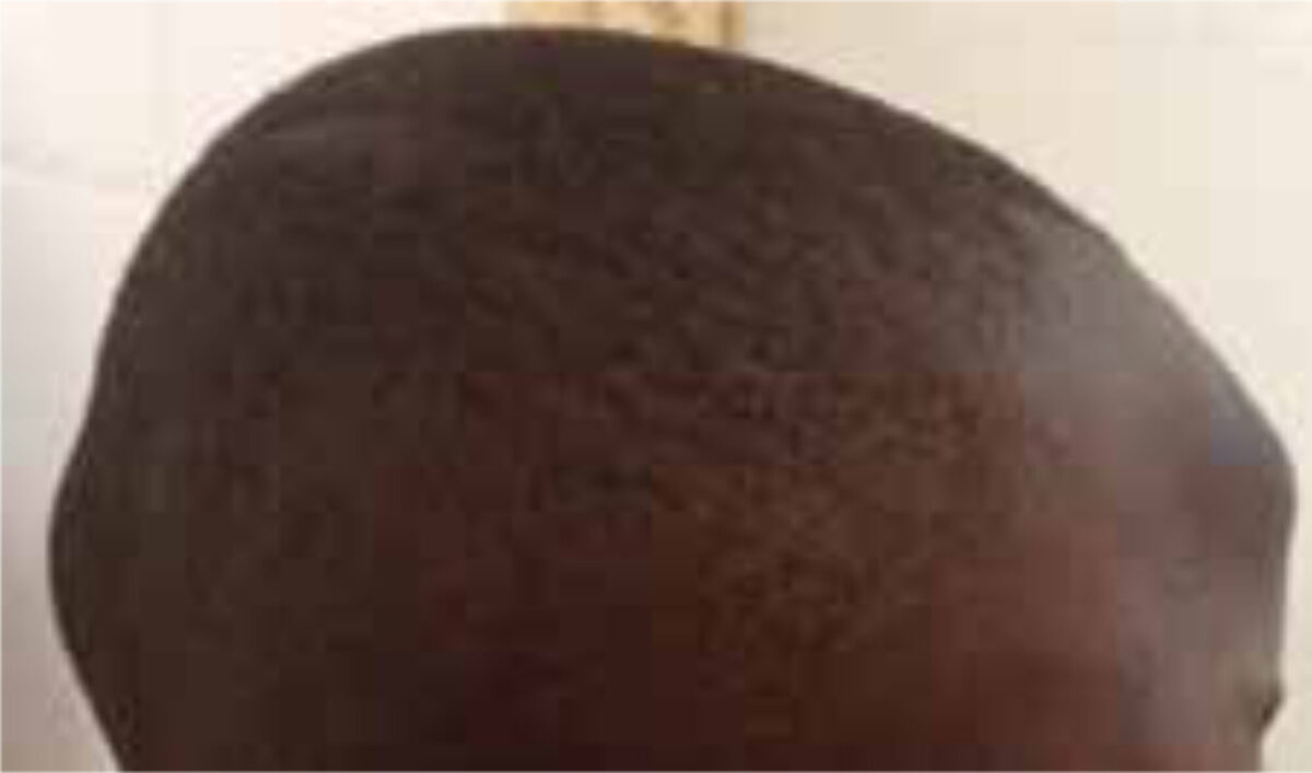
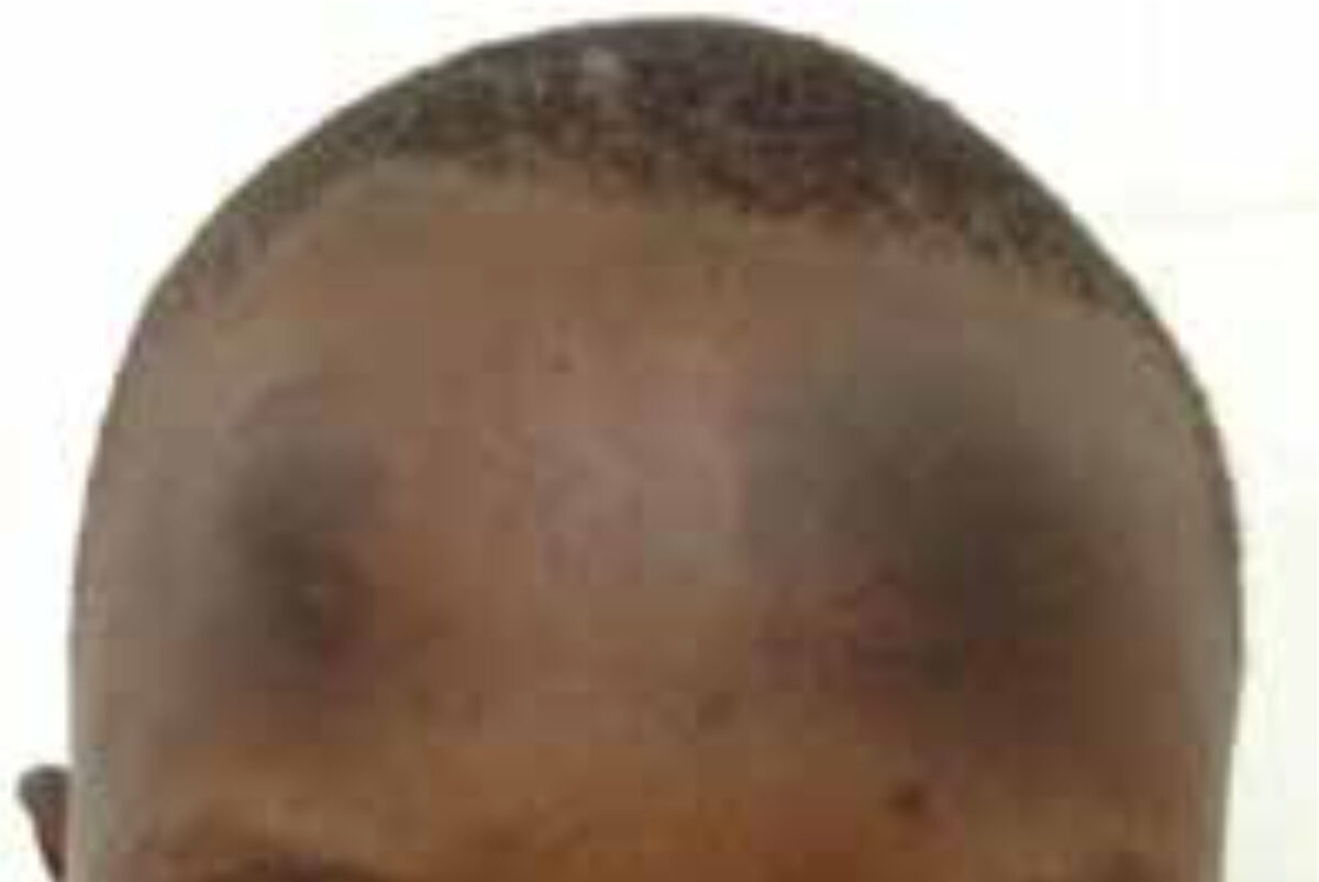
A 13-year-old boy presented at the out-patient department with two dark, elevated areas on the forehead (Figure 1 and 2). The onset was spontaneous without previous trauma. According to the patient and his parents, the discolouration occasionally resolved and returned. The lesions did not cause him any pain. The patient was convinced he was ‘strange’ and ‘different’, leading to absence from school. He had been treated with betamethasone cream without effect.
On physical examination, he was a healthy, well-nourished boy with two round, hyperpigmented masses on the forehead which were hard on palpation, giving the impression of bony tissue. The tumours were not attached to the skin and did not fluctuate. The skin was normal; there was no wound, lesion or scarring. There was no pain on palpation.
Specialist advice
The dermatologists and paediatricians were consulted regarding this case. Within two days, there were three replies with unanimous advice. The specialists suggested post-inflammatory hyperpigmentation as the underlying disorder, commonly caused by friction or rubbing. They proposed that nightly head banging (jactatio capitis nocturna) could lead to lesions in this particular area, by subconsciously hitting the head against a wall or bed during sleep. The tumours on the forehead could be hematomas, which may be hard on palpation. It was also proposed that, in case the patient was a Muslim, the lesions might be caused by repeatedly touching the floor with the forehead during prayer. They recommended taking a thorough history to ascertain the underlying inflammatory process.
Treatment
Unfortunately, no follow-up is available regarding this case as the patient did not return to the hospital for further treatment and evaluation. A further history as to whether this child was indeed a ‘head banger’ could therefore not be obtained.
Background of head banging
Jactatio capitis, or head banging, is part of a rhythmic movement disorder (RMD), which also includes headrolling, bodyrocking and bodyrolling.
Epidemiology
RMD is typically seen in the first year of life, resolves before the age of 10 and seldom continues into adulthood.[1] It more commonly affects males and is associated with ADHD, autism or mental retardation in older patients.[1,2] RMD can be aggravated during periods of stress and sleep deprivation.[3]
Clinical manifestations and diagnosis
RMD is defined by the International Classification of Sleep Disorders as repetitive, stereotyped movements of the large muscles, occurring anywhere in the body.[1] The movements last several minutes and typically occur during sleep onset and light non-REM sleep. Patients are often not aware of their condition.
Diagnosis
RMD can be diagnosed by observing nightly behaviour on video. More advanced diagnostic tools include polysomnography and videometry. As the differential diagnosis includes epilepsy, an EEG should be performed.
Prognosis
RMD is a self-limiting disorder and does not influence the development of children. However, adverse effects such as injuries, fractures and even subdural hematoma have been described.
Therapy
RMD does not need treatment apart from reassurance and patient education. Safety precautions and general sleep hygiene measures are recommended.[3] In severe cases, medical treatment with benzodiazepines can reduce the severity and frequency of RMD. Clonazepam has been reported to be effective.[4,5]
Background of postinflammatory hyperpigmentation
Postinflammatory hyperpigmentation (PIH) is a reactive hypermelanosis of the skin, occurring as a result of cutaneous inflammation.
Epidemiology
The exact prevalence of PIH is unknown. It can occur in any skin type but is most common in darker skin (Fitzpatrick skin type IV, V and VI: light brown to black).[6,7] It affects males and females equally and can occur at any age.
Etiology and pathophysiology
PIH can arise from different types of inflammatory processes, which can be divided into two categories: endogenous and exogenous conditions.[6,8] Among the endogenous causes are inherited diseases, cutaneous diseases, systemic diseases and allergic reactions. Exogenous conditions resulting in PIH include mechanical trauma, extremes of temperature, radiation, phototoxic reactions, chemical peels and laser procedures. Common causes include acne vulgaris, eczematous dermatoses and burn injury. In Muslims, a prayer mark can be considered.[9]
Two pathophysiologic mechanisms exist, leading to an excess or abnormal distribution of melanin either in the epidermis or in the dermis, thereby giving rise to two clinical subtypes: epidermal melanosis and dermal melanosis. PIH within the epidermis results from the release of inflammatory substances, stimulating melanocytes to increase production of melanin, with pigment being transferred to keratocytes. The dermal variant is caused by the release of melanin from basal keratinocytes, which have been damaged by an underlying inflammation process. The pigment is then phagocytosed by macrophages in the upper dermis.
Clinical manifestations and diagnosis
The affected areas correspond with the distribution of the initial inflammatory dermatosis.[6,7] Epidermal melanosis is characterized by light to dark brown lesions which are well-circumscribed. It may take 6-12 months to years to resolve without treatment. When the pigment is in the dermis (dermal melanosis), the lesions are darker blue-grey and poorly circumscribed. This hyperpigmentation can take years to fade to normal.[6]
PIH is a clinical diagnosis, based on the typical hyperpigmentation and a history of a preceding inflammatory process. A Wood’s lamp can be used to clinically distinguish dermal from epidermal melanosis. The diagnosis can be confirmed by biopsy, showing melanin in the dermis or epidermis.
Treatment
The first step in the treatment of PIH is eliminating the underlying inflammatory process. The patient should be advised to use high-factor sun protection and to avoid cosmetics, manual manipulation and harsh conditions (wind, cold, sunlight). Epidermal melanosis can be treated by topical depigmenting agents, retinoids, corticosteroids, chemical peels and laser modalities. Topical hydroquinone, which reduces melanin, is considered the gold standard therapy. Deeper pigmentation as in dermal melanosis does not respond well to the above mentioned treatment.[6] Patients may benefit from camouflage using cosmetic products.
References
- Lacz NL, Vafaie J, Kihiczak NI, Schwartz RA. Postinflammatory hyperpigmentation: a common but troubling condition. International Journal of Dermatology 2004;43:362-365.
- Davis EC, Callender VD. Postinflammatory hyperpigmentation. A review of the epidemiology, clinical features and treatment options in skin of color. The Journal of Clinical and Aesthetic Dermatology 2010;3(7):20-31.
- Saedi N, Dover JS, Ofori AO. Postinflammatory hyperpigmentation. UpToDate 2015.
- Manni R, Terzaghi M. Rhythmic movements during sleep: a physiological and pathological profile. Neurological Sciences 2005;26 Suppl 3:s181-185.
- Stepanova I, Nevsimalova S, Hanusova J. Rhythmic movement disorder in sleep persisting into childhood and adulthood. Sleep 2005;28(7):851-857.
- Wills L, Garcia J. Parasomnias: epidemiology and management. CNS Drugs. 2002;16(12):803-810.
- Chisholm T, Morehouse RL. Adult headbanging: sleep studies and treatment. Sleep 1996;19(4):343-346.
- Hashizume Y, Yoshijima H, Uchimura N, Maeda H. Case of head banging that continued to adolescence. Psychiatry and Clinical Neurosciences 2002;56(3):255-256.
- Orenay OM, Sarifakioglu E. Prayer Mark on the Forehead: Hyperpigmentation. Annals of Dermatology. 2015;27(1):107-108.