Main content
Setting
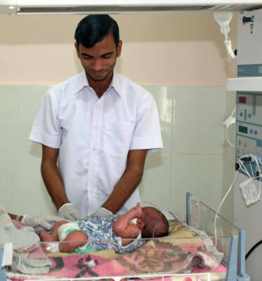
We report two cases from a rural health clinic in Namatanai, in the New Ireland province of Papua New Guinea. The clinic has an inpatient ward with 40 beds, very basic diagnostic facilities and a small operating theatre. The nearest referral hospital is a five hour drive. During rounds, two patients with a similar skin eruption caught the attention of the doctor in charge.
Case 1
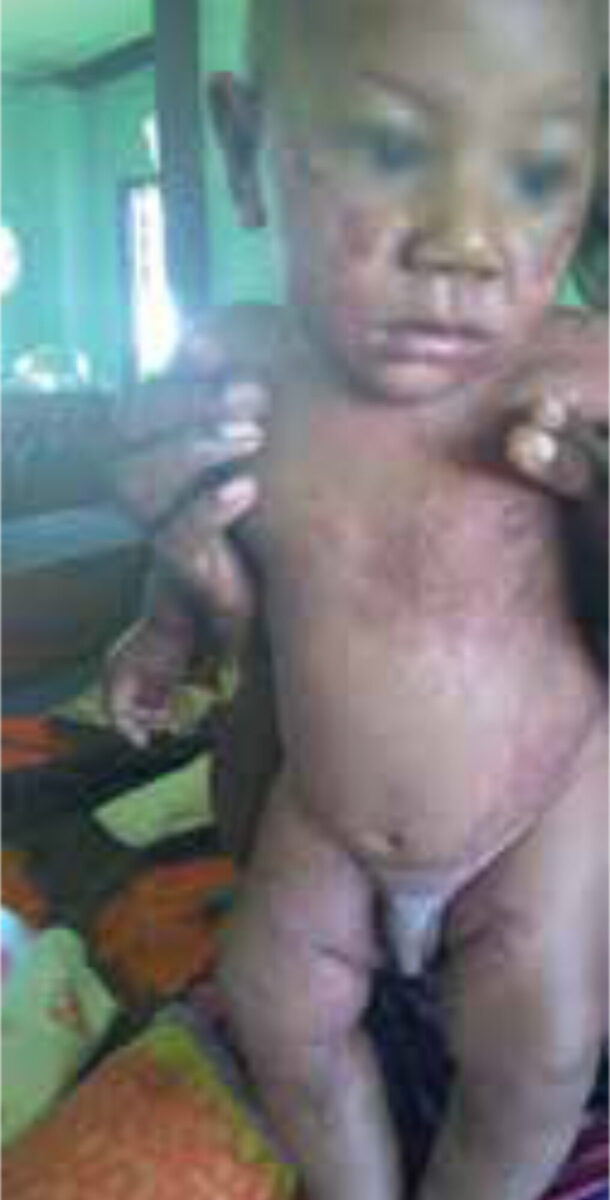
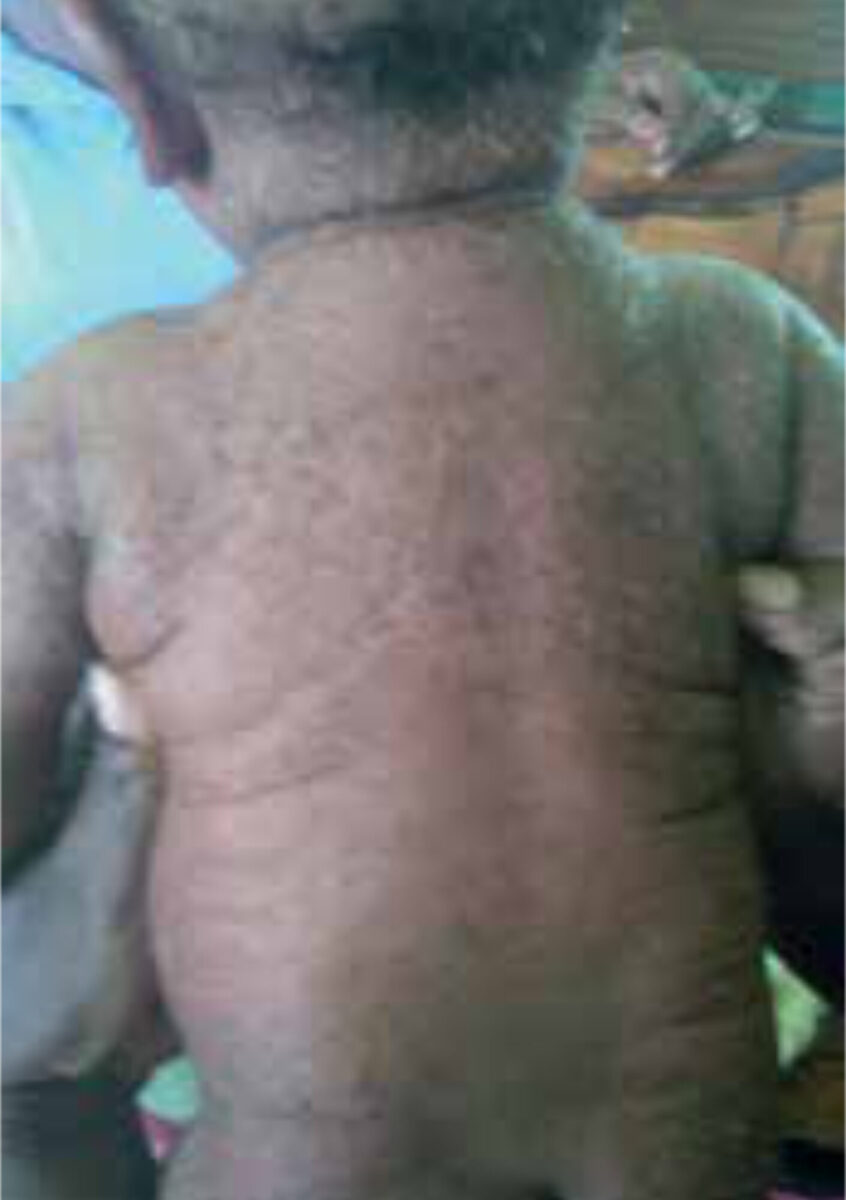
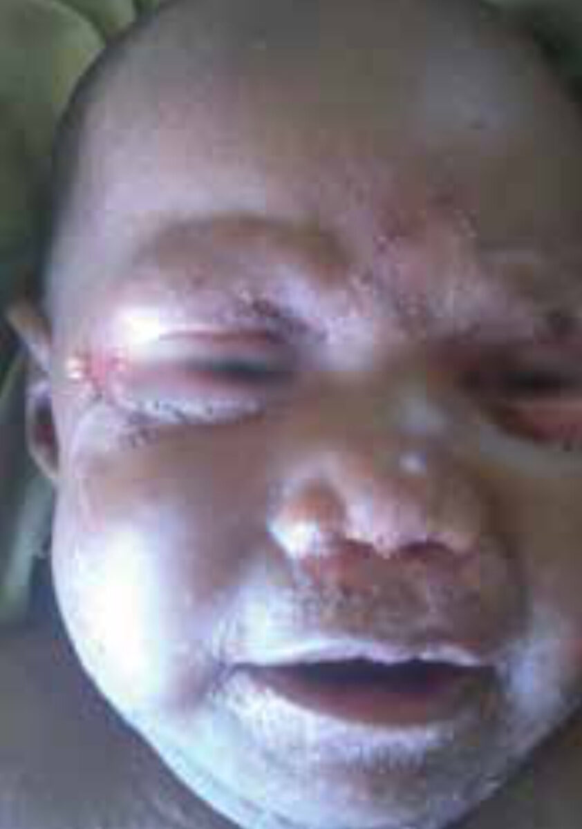
A male infant, 2 months old, had presented at the outpatient department with stomatitis and diffuse erythema. It had started 6 weeks before as a facial rash, but had disseminated to cover the whole body. On examination, symmetrical erythematous patches and plaques were observed, mainly in the body folds and facial area. The child seemed uncomfortable on touching the lesions. The mother had been using 5 different creams (mostly local herbs and plants) without success.
Both the patient and his mother tested positive for syphilis (using the VDRL test), for which they were treated. Following admission in the hospital and treatment with oral flucloxacillin, intravenous hydrocortisone and betamethasone cream, the eruption seemed to improve. However, after a while, the extent of the rash was found to increase.

Case 2
The second case was similar, concerning a 4 month old girl. She presented with conjunctivitis and an acute rash in the diaper area, which had spread to the trunk and face within a week. There were erythematous papules on the trunk, along with periorbital, perinasal and perioral patches and plaques. As in case 1, the eruption seemed to arise particularly in the body folds. The child was irritable on examination. The patient had tested negative for syphilis, contrary to her mother. She had been hospitalized for 2 days and had been treated with oral flucloxacillin, betamethasone cream and ophthalmic tetracycline. To date, there had been no improvement. Both cases were term infants with unknown birth weight; both were receiving breast milk. There was no evidence of growth impairment, malnourishment, diarrhea, alopecia or recurrent infections.
Specialist advice
The dermatologists were asked for advice on diagnosis, treatment and prognosis. Three dermatologists responded to this enquiry.
Their differential diagnosis included (seborrheic) eczema, allergic reaction to local ointments, intertrigo, malnutrition, and zinc deficiency. The advice was to continue betamethasone cream (diluted with vegetable oil, as betamethasone alone would be too strong for these young children) and to start zinc supplementation. The lesions were not found to be typical for syphilis. Nevertheless, the advice was to treat, as neonatal syphilis cannot be excluded simply by negative testing.
Treatment
Both children were started on zinc supplementation (zinc sulfate, tablets, 10mg daily). Betamethasone cream was diluted and continued. After 2-5 days, both patients recovered completely.
Background of zinc deficiency
Zinc metabolism
Zinc status depends on several factors, including bodily stores, dietary intake, absorption and endogenous losses [1]. Zinc is found in animal products such as meat and milk [2]. 30% of ingested zinc is absorbed by the duodenum and proximal small intestine [2,3]. Zinc is secreted from the body mainly by the gastrointestinal system and the kidneys.
Etiology of zinc deficiency
Zinc is an essential mineral. It plays a role in protein, carbohydrate and vitamin A metabolism and is needed for growth, development, cell proliferation, healing and tissue repair [2-4]. The recommended daily dosage of zinc varies according to age. 17% of the world population is estimated to have a low dietary intake of zinc, particularly in developing countries [3].
Acquired zinc deficiency is principally seen in breastfed infants. Zinc content of breast milk physiologically declines in the first months postpartum, from >3mg/L to <1mg/L after 6 months [1]. Therefore, the infant will need other sources of zinc (e.g. complementary foods) to meet daily requirements. Particularly prone to zinc deficiency are premature infants, as they have smaller liver zinc stores, lower absorptive capacity and larger intestinal and renal losses. Other risk factors for acquired zinc deficiency are diseases with decreased absorption and increased secretion of zinc, such as celiac disease, cystic fibrosis, short gut syndrome and liver disease.
The most important congenital form of zinc deficiency is acrodermatitis enteropathica (AE). It is an autosomal recessive disorder, resulting from a partial block in zinc absorption in the intestines [1]. Several genetic mutations have been found to cause AE. Symptoms typically commence after the infant is weaned off the breast, as absorption of zinc is decreased in bovine milk as compared to human breast milk [4,5].
Another genetically based zinc deficiency has a maternal lactogenic origin [1]. Mutations in zinc transporters localized in the mammary glands lead to an inability to secrete zinc in breast milk. Therefore, low zinc concentrations are found in breast milk despite a normal maternal zinc status [1].
Clinical features and diagnosis
Acquired and congenital forms of zinc deficiency are clinically indistinguishable [5]. They are characterized by growth impairment, loss of appetite and recurrent infections (due to immune deficiency) [1]. The classic triad of AE consists of diahrroea, alopecia and acral and periorifical dermatitis, but this triad is only present in 20% of all patients [2]. Skin disorders include eczematous or erythematous lesions and ulcerations, predominantly occurring periorally, periorbitally, in diaper regions and on the extremities. Paronychia, onycholysis, stomatitis and conjunctivitis may be found. Other clinical features include apathy, irritability, photophobia and hypogonadism [2].
Determining zinc levels in serum remains the gold standard for diagnosing zinc deficiency. However, this biomarker has several limitations. It fluctuates with meals and time of day, has low sensitivity for detecting marginal zinc status, acts as a negative acute-phase reactant to inflammation, and samples can be contaminated by skin or collection materials [1]. Therefore, recognizing clinical features and taking a thorough history (including feeding history and potential gastrointestinal pathology) is imperative.
Treatment
Oral administration of zinc is the treatment of choice, regardless of the origin of zinc deficiency. Zinc sulfate is best tolerated, although zinc can also be administered as acetate, amino acid chelates and gluconate [2]. An initial dose of 5-10mg/kg/day is usually recommended, followed by a maintenance dose of 1-2mg/kg/day [2]. Dosage should be increased during pregnancy and adolescence [2].
Duration of treatment depends on clinical response and pathophysiology. Supplementation can be discontinued after resolution of symptoms, but should be lifelong in patients with AE. The effect of zinc administration will become apparent after 24-48 hours, with skin disorders improving and resolving completely within 2-4 weeks [2,4].
Adverse effects of zinc are rarely seen with small dosage and short treatment. However, with prolonged duration of treatment, gastro-intestinal symptoms such as nausea, vomiting and hemorrhage can arise [2]. Furthermore, high serum levels of zinc can result in hypocupremia, warranting monitoring of copper levels throughout treatment.
Take-home messages
The tropical doctor in charge formulated three lessons from these cases. Start zinc supplementation in any child with a skin disorder. Use vegetable oil to dilute betamethasone for small children. Ask mothers about local ointments and treatment previously used.
References
- Krebs NF. Update on zinc deficiency and excess in clinical pediatric practice. Ann Nutr Metab 2013;62(suppl 1):19-29.
- Perafán-Riveros C, França LF, Alves AC, Sanches JA Jr. Acrodermatitis enteropathica: case report and review of the literature. Pediatr Dermatol 2002;19(5):426-431.
- Ogawa Y, Kawamura T, Shimada S. Zinc and skin biology. Arch Biochem Biophys 2016; http://dx.doi.org/10.1016/j.abb.2016.06.003.
- Haliasos EC, Litwack P, Kristal L, Chawla A. Acquired zinc deficiency in full-term newborns from decreased zinc content in breast milk. Cutis 2007;79:425-428.
- Zattra E, Belloni Fortina A. Transient symptomatic zinc deficiency resembling acrodermatitis enteropathica in a breast-fed premature infant: case report and brief review of the literature.
- G Ital Dermatol Venereol 2013;148(6):699-702.