Main content
Setting
Bale Robe is situated in south-central Ethiopia in the Oromia region, approximately 430 kilometers from the capital, Addis Ababa. This region is known for its fertile highlands used for agriculture and livestock breeding. The Bale Robe District hospital has an annual catchment population of approximately one million people with a low HIV prevalence of 2 to 3 percent. The medical staff exist of one surgeon, one gynaecologist, one tropical doctor and nine general practitioners. The closest referral hospital is five hours’ travelling from Bale Robe.
Case
A 34-week-pregnant HIV uninfected woman, gravida 2 para 1, was referred to the Bale Robe Hospital in Ethiopia because of severe pre-eclampsia. Despite treatment with magnesium sulfate, hydralazine and dexamethasone, the high blood pressure persisted and ascites developed. Subsequently, labour was induced by priming with misoprostol and a girl was delivered without complications or signs of fetal distress. The birth weight was 2200 grams and APGAR scores at 1 and 5 minutes were 8 and 9, respectively. There were no signs of infection in the newborn infant.
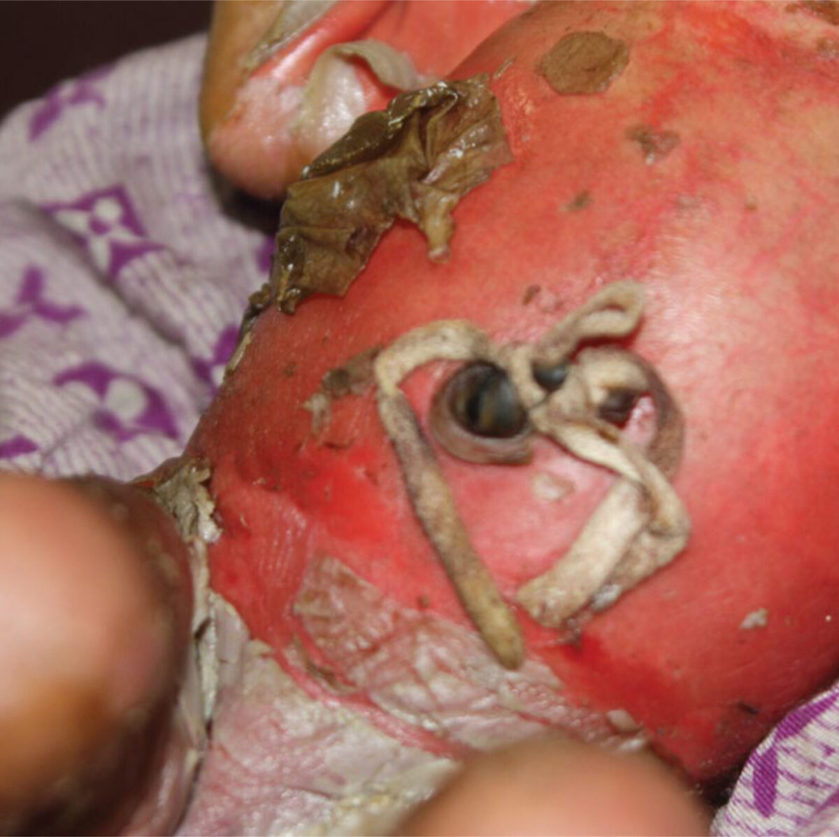
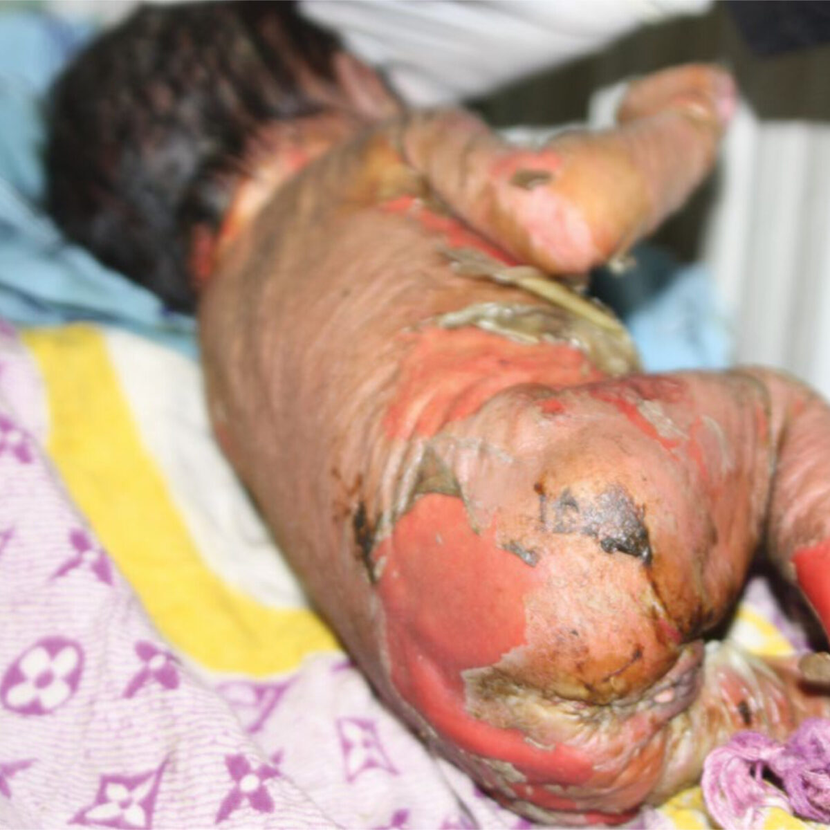
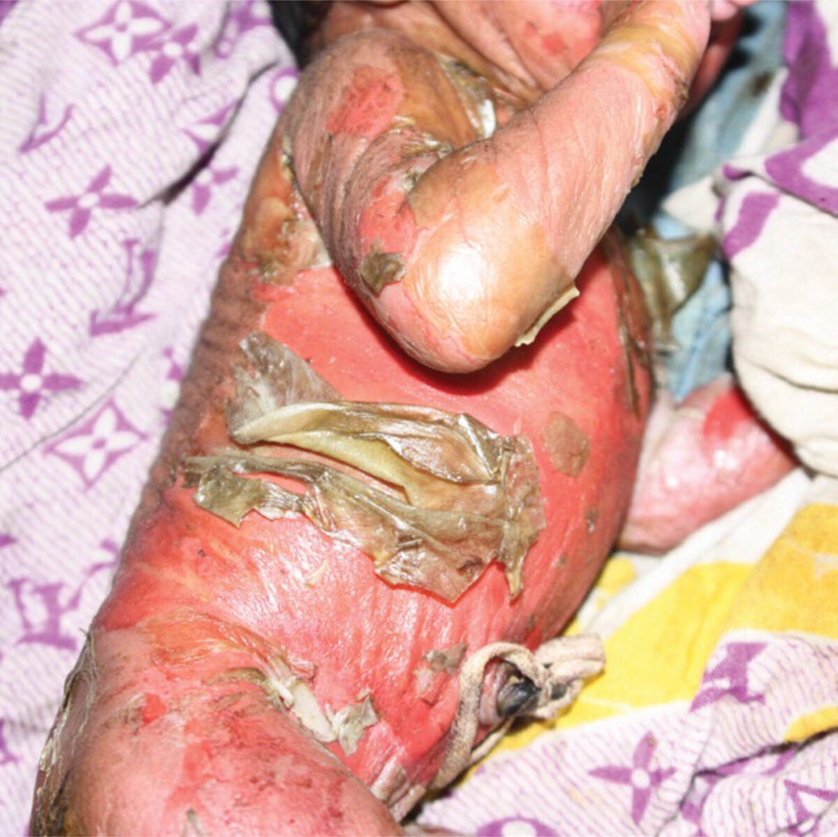
The midwife reported a white translucent skin, appearing as a membrane covering the infant. Immediately after birth the membrane detached and blisters developed all over the body surface. Two days later the membrane was dried, wrinkled, gray and it peeled off from most of the skin surface and at these sites a moist, red skin was observed. The trunk and extremities were mostly affected. The head, face, mouth and genitals were relatively unaffected. The infant cried forcefully and appeared to be in severe pain when manipulated. Further physical examination showed no other abnormalities and no fever.
The pediatricians and dermatologists were asked for their advice via Consult Online.
Advice from specialists
Within a matter of hours the dedicated specialists gave their opinion, resulting in a vivid discussion about the possible illness of this child. Several diagnoses were suggested.
Collodion baby syndrome
The diagnosis first emerging from their e-mails was the Collodion baby syndrome due to the translucent so-called “collodion” membrane covering the infant’s body. This is an uncommon clinical presentation of several genetic conditions resulting in abnormal cornification of the skin. Most patients are ultimately given the diagnosis lamellar ichthyosis or congenital ichthyosiform erythroderma characterized by onward skin thickening due to hyperkeratosis. As patients age the scale will become thicker mainly on the soles of hands, feet en flexion areas.
In this child, however, the blisters were a predominant feature, as can be seen in the pictures. Therefore the rare condition congenital bullous ichthyosiform erythroderma was suspected, which is characterized by collodion membrane followed by widespread blisters and erosions. Several mutations in the many keratin genes have been associated with this condition.
The main challenge in all infants with Collodion baby syndrome is the immediate risk of dehydration and infection due to an impaired skin barrier. [1,2]
Epidermolysis bullosa
An important differential diagnosis was Epidermolysis bullosa, characterized by generalized blistering and erosions induced by minor trauma, caused by a number of specific mutations in the keratin genes. Severe forms present at birth with extensive blistering; milder variants can be detected in the first years of life or even in adulthood. [3]
Staphylococcal scalded skin syndrome
Although in this case the first symptoms were present at birth and no fever was detected, an infectious cause was also considered. The severe Staphylococcal scalded skin syndrome (SSSS) is known to cause generalized blistering due to toxins produced by S. Aureus. Typically, presentation occurs at 3 to 7 days of age with a generalized erythema beginning around the mouth. Flaccid blisters arise one day later, predominantly affecting skin areas exposed to mechanical stress. Treatment consists of immediate antibiotic therapy. [4]
Toxic epidermal necrolysis
TEN is considered the more severe variant of Stevens-Johnson’s syndrome and is characterized by necrosis of the epidermis and mucous membrane involvement induced by a variety of drugs including sulfonamides, anticonvulsants and NSAIDs. Clinically erythematous skin lesions with a darker centre arise evolving into vesicles and bullae. It may be difficult to distinguish from SSSS.
TEN is a very rare but fatal condition in early infancy, only a few cases have been reported, all after administration of drugs to the child itself. [5]
Discussion
The dermatologists and pediatricians agreed that based on clinical presentation the Collodion baby syndrome was the most probable diagnosis, presumably caused by congenital bullous ichthyosiform erythroderma. There is also a fair possibility the infant suffered from Epidermolysis bullosa. It would have been helpful to distinguish from this condition by rubbing the skin and to observe whether a blister would arise. Unfortunately it was not possible to carry out this test due to a delay in presentation and correspondence with the specialists.
As mentioned before an infectious cause like SSSS is less likely because the child presented with skin abnormalities at birth and did not have a fever. Obviously an infection would at least need a few days for incubation.
The remaining condition, TEN, is less likely because no drugs had been given to the infant prior to the development of skin abnormalities.
A skin biopsy would have given the ultimate confirmation of the diagnosis, but as in many district hospitals a lack of resources impaired proper histopathological analysis. Despite the lack of a precise diagnosis all specialists underlined the dehydration and infection risks of a major skin defect. Therefore they advised adequate hydration, systemic antibiotic treatment and topical treatment for the skin defects as if they were burns.
Follow-up
The infant was placed into kangaroo mother care. Although the mother was very anxious when manipulating her child she did a good job with breast-feeding and keeping the child close to her chest. The child urinated regularly so dehydration did not seem to be a major problem. Because of a high infection risk the infant was treated with gentamycin, ampicillin and topical nitrofuzone.
The following days the skin condition did not change. The child gave the impression of being in a lot of pain and nothing but paracetamol was available. Unfortunately the child died several days later.
Acknowledgment
We kindly thank dr Ben Naafs for his useful comments and advice.
References
- Collodion baby: an update with a focus on practical management. Prado R, Lixia E, Gambe R et al. J Am Acad Dermatol 2012;67:1362-74.
- Overview of the inherited ichthyoses. Choate K, Dyer J, Corona R et al. Up to date 2014.
- Epidemiology, pathogenesis, and clinical features of epidermolysis bullosa. Laimer M, Dyer J, Corona R et al. Up to date 2014.
- Vesiculobullous and pustular lesions in the newborn. Pielop J, Levy M, Corona M et al. Up to date 2014.
- Toxic epidermal necrolysis in early infancy. Scully MC, Frieden IJ. J Am Acad Dermatol. 1992 Aug;27(2 Pt 2):340-4.