Main content
Setting
This case is from Nyeri, in the South of the Kati province, central Kenya. A team of the Dutch non-profit organization Medical Checks for Children (MCC) is working there on a paediatric project. Children of different schools and orphanages in the area are screened for medical problems. A regular physical examination is performed and there is a possibility for rapid haemoglobin, malaria and urine tests. For additional laboratory tests, imaging or advanced treatment, the patient must be referred to a hospital in the area. Often patients do not have the financial means to pay for this themselves. In some cases, financial support can be provided by a partner organization of MCC.
Case
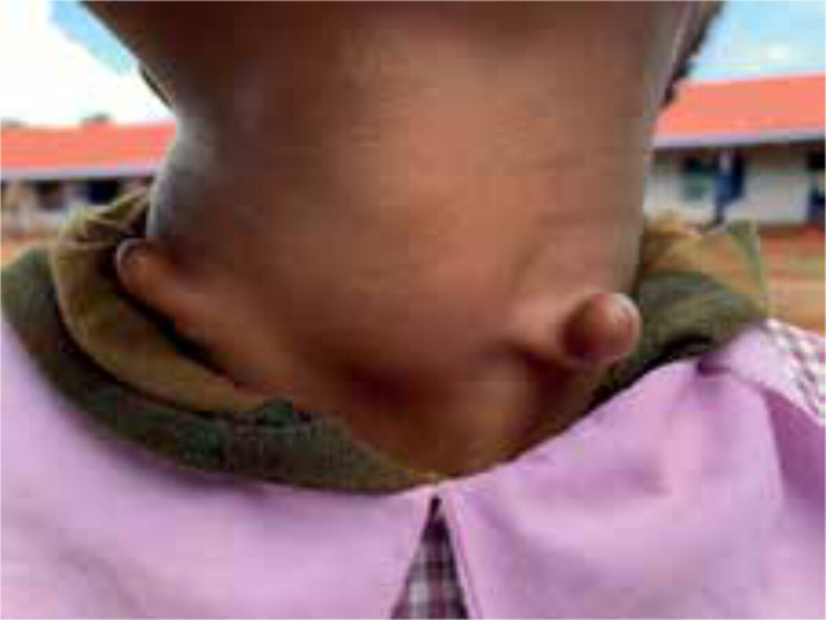
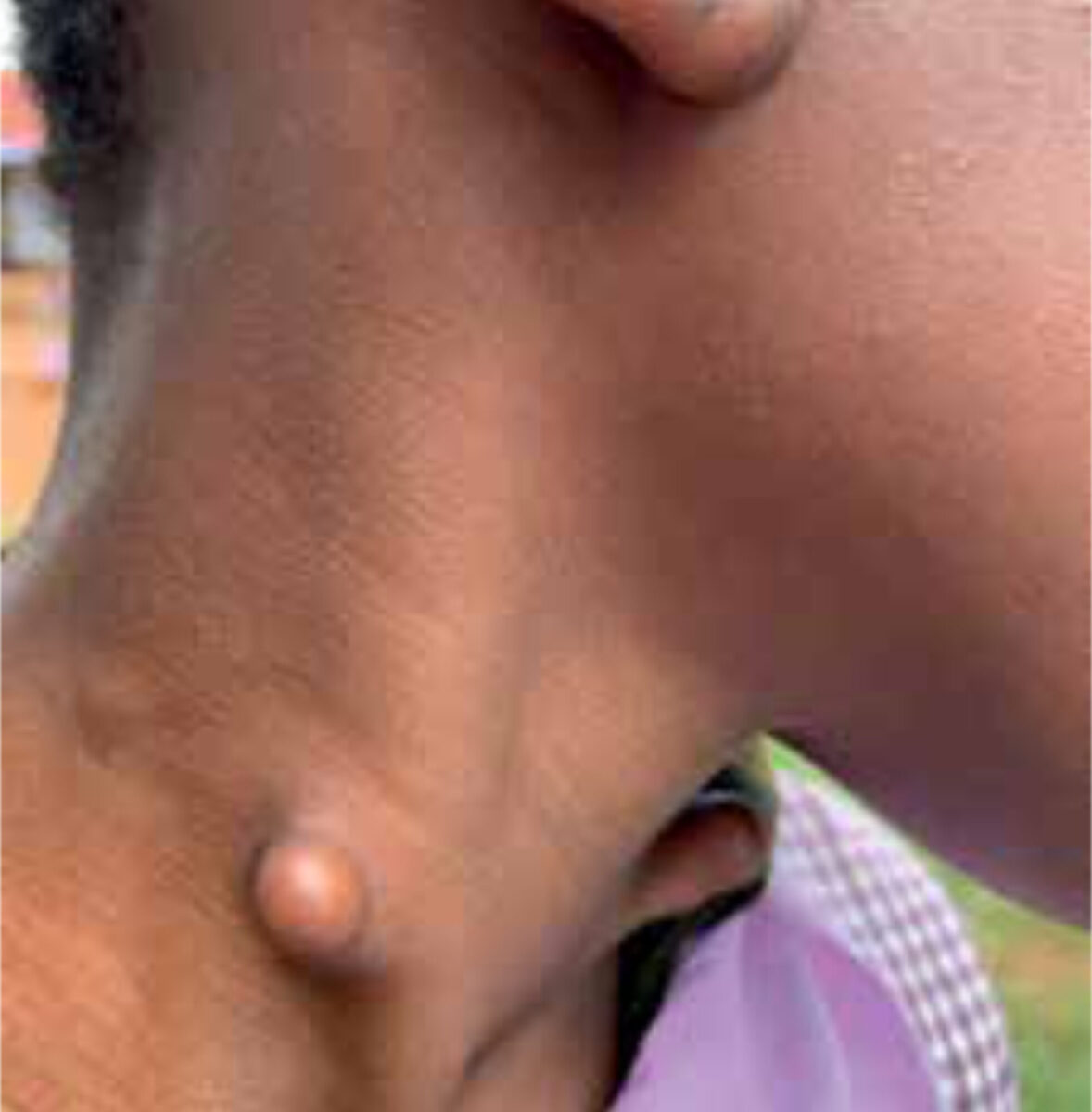
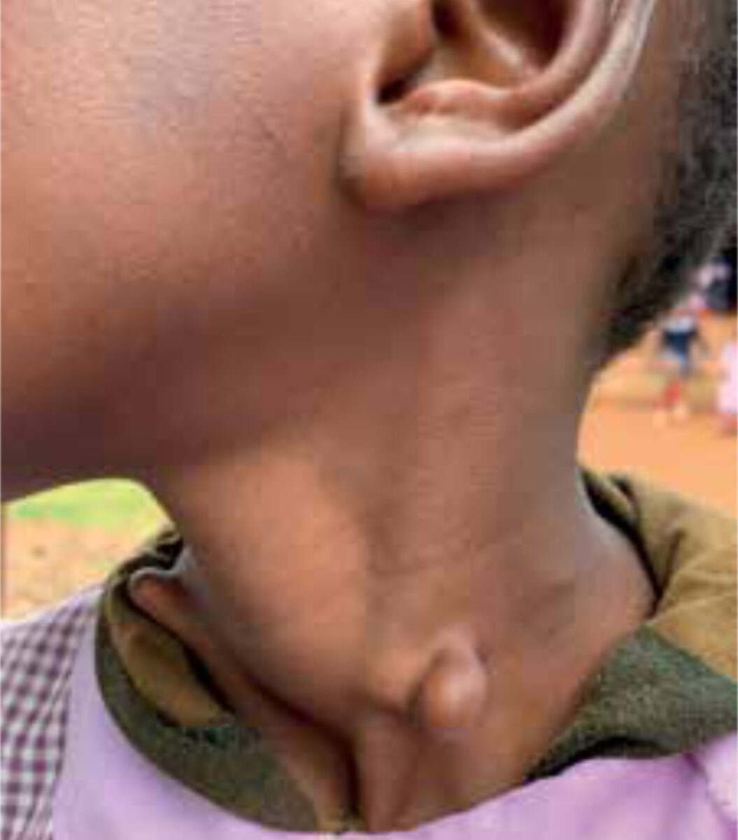
A 9-year-old girl presents with a swelling on both sides of the neck (Figures 1-3). The swellings have been there for as long as she can remember and have grown proportionally with her body growth. Except for the cosmetic appearance, she does not have any other complaints. On clinical examination, two fixed elastic tags are palpable, and the lateral side of the left tag has a small hardening. Palpation is not painful. Her voice sounds normal while talking, and she does not experience any problems swallowing food. Heart sounds are normal. The remainder of the general physical examination shows no abnormalities. The haemoglobin level is normal. No additional tests were performed.
Specialist advice
The paediatrician thought of cervical chondrocutaneous branchial remnants. An ultrasound could support this diagnosis. A consultation of a paediatric surgeon would be advisable to discuss the possibility of removal.
Cervical chondrocutaneous branchial remnants
Cervical chondrocutaneous branchial remnants (CCBR) are rare, benign, congenital malformations that are present at birth.[1] It is most often located in the middle or inferior anterolateral part of the neck, anterior to the sternocleidomastoid muscle.[2,3] Although uncommon, chondrocutaneous branchial remnants can be present at other locations, including the oral cavity, nasopharynx, middle ear and eyes.[1] Due to its rareness, little is known about these malformations, and literature mostly consists of case reports. Until 2020, only 117 cases had been described in medical literature.[3]
CCBR is a form of choristoma, a histological term for a benign tumorous mass of developmental origin that consists of normal body tissue localized at an abnormal body site.[1] A CCBR mass contains an elastic or hyaline cartilage core, covered by subcutaneous fatty tissue and normal skin (dermis and epidermis).[2,3] It usually presents as a solitary mass and has no connection to surrounding tissue but can be adhered to the fascia of the sternocleidomastoid muscle.[3] It is not associated with underlying cysts.[3] Diagnostic imaging by ultrasound or CT scan can support the diagnosis of CCBR, typically showing a tubular shaped cartilage core extending to the sternocleidomastoid muscle and surrounded by fatty and normal skin tissue.[2] Histological analysis can confirm the diagnosis.[3]
Although the exact origin of CCBR is not yet known, it has been established that it is an embryological remnant that most likely arises from incomplete disappearance of the embryonic pharyngeal arches, leaving cells that differentiate into cartilage at an abnormal body site.[3]
The embryonic pharyngeal arches (also known as branchial arches) start to develop in the fourth week of pregnancy and are precursors for many structures in the neck, face and head of the developing embryo.[4] The first and second branchial arch are, among other things, responsible for the development of the auricle.[1,4] In the sixth week, these arches form auricular hillocks (precursors for the outer ear) that, in the seventh week, migrate from the inferior part of the anterolateral neck to the lateral side of the head.[1,4] The second and third arches form, among others, the hyoid bone of the larynx.[4] The fourth and sixth arches form the cartilage of the larynx.[4]
| Thymic cyst |
| Thyroglossal duct cyst |
| Branchial cleft cyst |
| Hair follicle naevi |
| Congenital hamartomas |
| Fibroepithelial polyp |
| (Epi)dermoid cyst |
| Squamous papillomas |
| Teratoma |
| Embryoma |
| Choristoma |
Two theories exist about the origin of these malformations.[1] The first one claims that CCBR originates from auricular tissue due to problems with the migrating process. The second one claims that it derives from the laryngeal remnant of the lower pharyngeal arches.[1] The presence of elastic cartilage in CCBR therefore implies an auricular origin from the first or second pharyngeal arch, while the presence of hyaline cartilage in CCBR implies an origin of remnants from the second or lower arches.[1,2,3]
CCBR grows (very) slowly or not at all.[3] It seems predominant in males and can present as bi- or unilateral, the latter being the most common appearance. Familial forms have been reported.[1,2,3] CCBR generally presents with normal skin and no signs of inflammation or discharge.[1,3] The differential diagnoses of masses in the neck of a new-born can be broad (Table 1).
Treatment of CCBR is mostly for cosmetic reasons since it is benign and shows little to no growth. It is important to keep in mind that it can have a negative psychological impact on a person’s life.[3]. Recommended treatment is early and complete surgical excision for the best cosmetic results.[1,2,3] However, surgical treatment can be postponed to a suitable time and place later in life.[3]. It is advisable to have histological confirmation of the tissue after removal.[2,3]
Associated anomalies are common (11%-76%) with CCBR (Table 1). These most often present in the auditory, cardiovascular, or genitourinary tract (Table 2).[1,3] Evaluation for potential anomalies is therefore advised via a full physical examination and (preoperative) imaging of the heart and abdomen.[2,3]
SYSTEM ANOMALY Auditory neurosensory deafness, serous otitis media, malformation of external ear Respiratory Tracheomalacia Oral-gastrointestinal Cleft palate, oronasal reflux, inguinal hernia Genitourinary Hydronephrosis Cardiovascular Atrial septal defect Musculoskeletal – Visual –
Follow-up
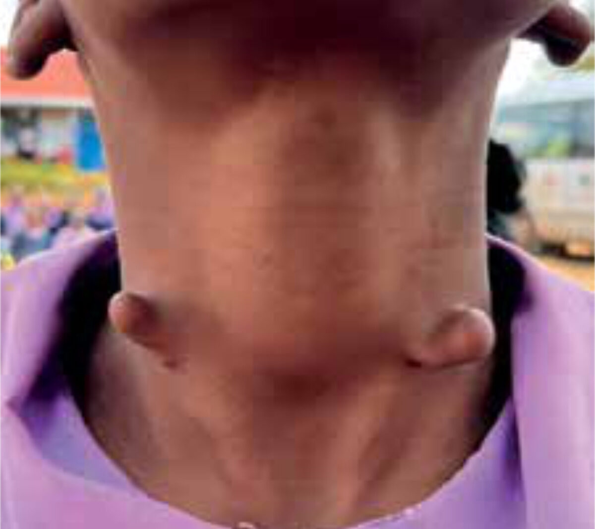
The diagnosis in this patient was made through a combination of a detailed history (present since neonatal period, stable size, no physical complaints) and a thorough physical examination. Because of referral problems during the covid pandemic and the absence of direct clinical consequences, an additional ultrasound for the masses was not effectuated. The patient does not experience any psychological problems because of the malformation. This, in combination with the absence of any physical need for intervention and the relatively high costs for removal, made the choice for a conservative approach in this setting preferable. She is being followed up every year (Figure 4), and the masses show no growth. If problems present in the future, she might need to be referred to the hospital for additional tests or surgery.
References
- Raed Al-Taher, Marzouq Amarin, Zakaria Shkoukani, Ahmad Mansour, Nader M. Albsoul. Cervical chondrocutaneous branchial remnant. Journal of Pediatric Surgery Case Reports 2017 Jul;25:31-33
- Ginat DT, Johnson DN, Shogan A, Cipriani NA. Cervical Chondrocutaneous Branchial Remnants. Head Neck Pathol. 2018 Jun;12(2):244-246.
- Lee HS, Kim TH, Jang JY, Woo JW, Lee J, Jeong SH, Jung EJ, An HJ, Park T. Bilateral cervical chondrocutaneous branchial remnants: A case report and a review of the literature. Medicine (Baltimore). 2020 Jul 10;99(28):e21114.
- Moore KL, Persaud TVN, Torchia MG. The Developing Human: Clinically Oriented Embryology. 8th edition. Philadelphia: Saunders, Elsevier; 2008.