
Main content
Setting
The LAMB hospital (Lutherian Aid to Medicine Ban-gladesh) is located in the Dinajpur District in the Northwestern part of Bangladesh, approximately 320 kilometers from Dhaka. It is a general hospital with a capacity of 150 beds and a large outpatient clinic run by 200 staff members, mainly Bangla-deshi with a small number of expatriates. Annually, healthcare is provided to 10,000 inpatients and 6,000 outpatients. The population of Bangladesh is highly dense and fast growing and natural disasters occur frequently.
Case
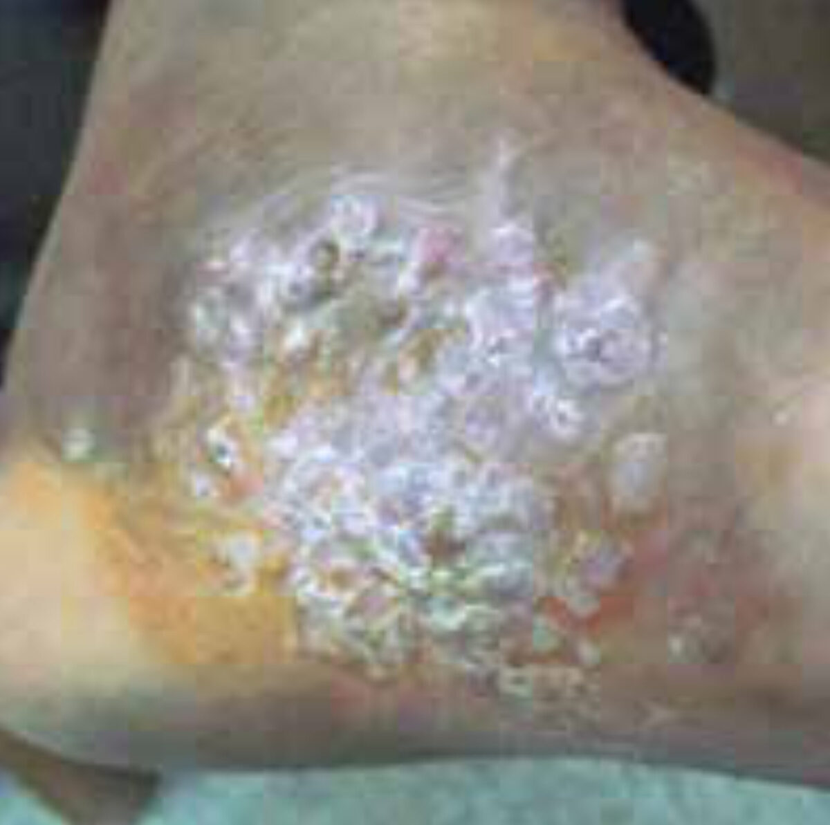
A 30-year-old male complained of a painful and slow growing swelling of his left foot and difficulty in walking. The condition started two years ago when he had a splinter removed. Upon examination at the medial side of the left foot, a swelling of 60 millimeter in diameter was seen with multiple sinuses, continuing in the deeper tissues, which was non-mobile and painful (Figure 1). The patient men-tioned some discharge from the lesion. A biopsy was performed in another hospital and histopathology showed acute and chronic inflammation without signs of granuloma, malignancy or fungal infec-tion. The attending surgeon considered an amputation but first consulted the Consult Online surgeons for their expertise on this matter.
Specialist advice
Within one day two surgeons gave their opinion and advice on this case. Both suspected a Myceto-ma, until recently also known as Madura foot, due to the history and the presence of sinuses. They suggested that the biopsy may have been too superficial to lead to a conclu-sive diagnosis.
An extensive wound debridement and simultaneous deep tissue biopsy was ad-vised. They also rec-ommended taking X-rays of the affected body part to look for additional bone infection.
Treatment and follow up
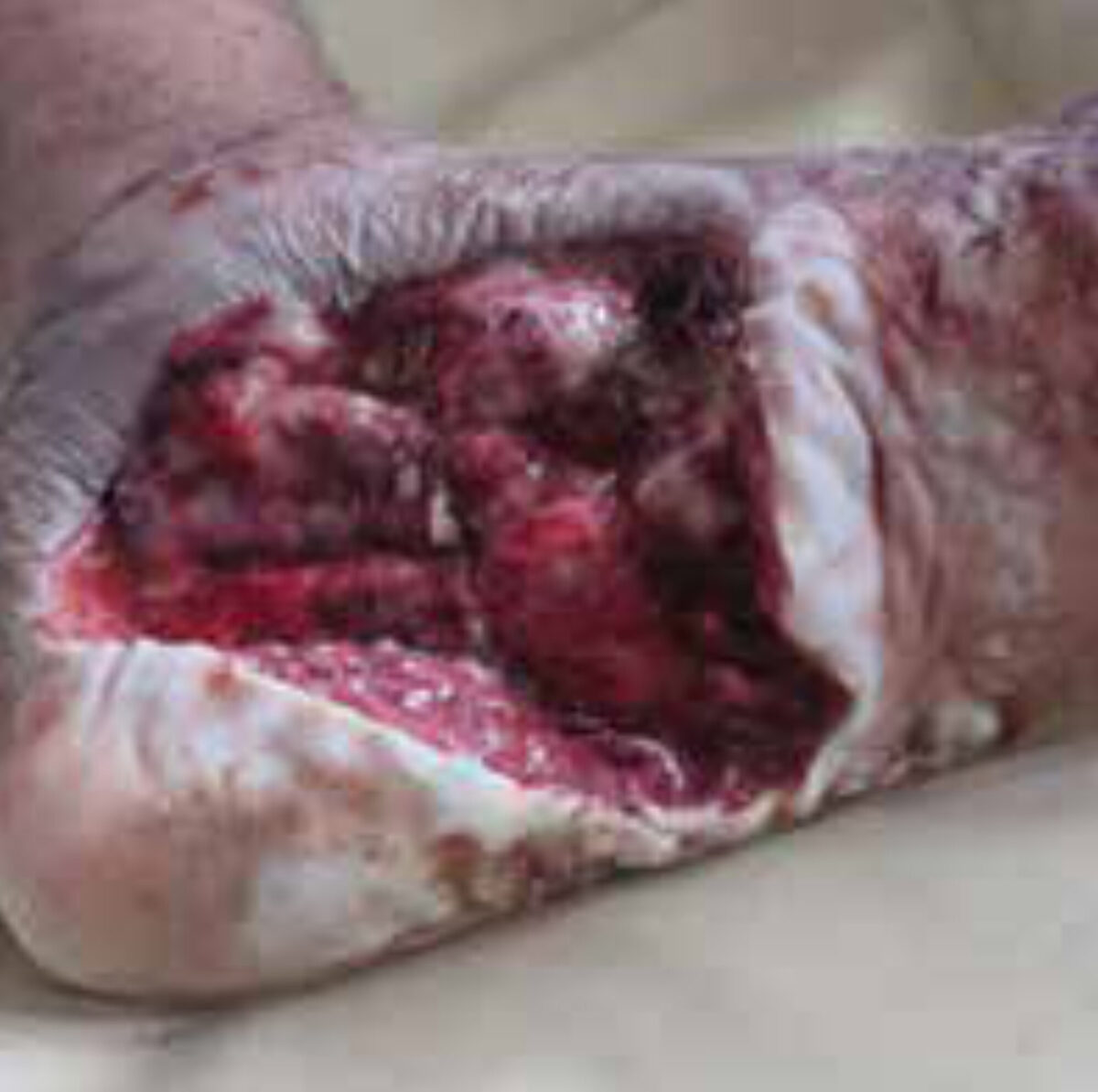
A plain X-ray of the left foot showed no signs of bone involvement. Consistent with the given advice a wound debride-ment and a deep tissue biopsy were carried out. Figure 2 shows the post-operative result. The patient was also treated with griseovulfine, a fungistatic antibiotic. Itraconazole or keto-conazole would have been the drug of first choice, but it would be prohibitively expensive to use for a long period of time.
Eighteen days after initial surgery the wound appeared infected. A culture revealed a Staphylococcus aureus infection. Cloxacillin was added to the treatment regime. In the meantime the result of the deep tissue biopsy indicated the presence of colonies of Actinomy-cetes species within acute inflam-matory cells. This confirmed the suspected diagnosis. Ceftriaxone and later cotri-moxazole were add-ed to the treatment regimen.
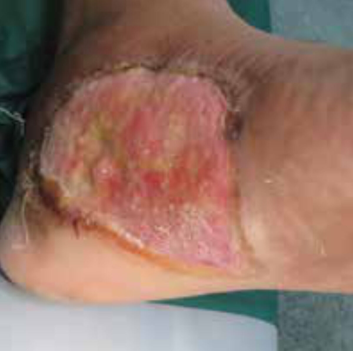
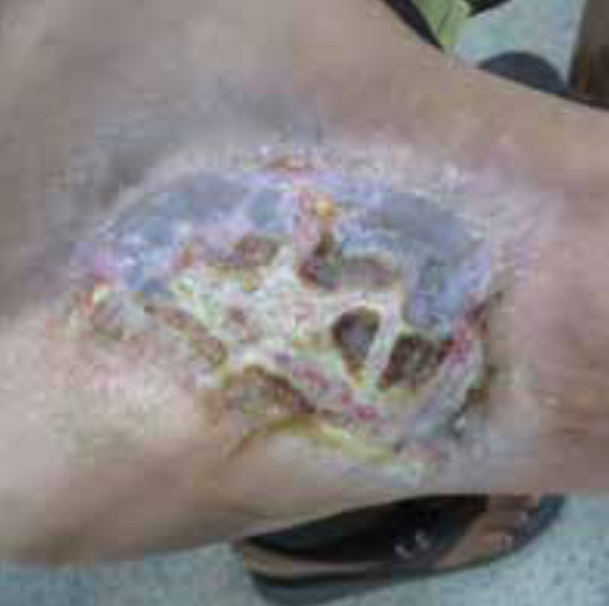
After 2,5 months of treatment the wound was covered with granulation tissue and showed no signs of infection (Figure 3). During a second operation a split skin graft was done and after I month the wound was almost entirely covered with normal skin, as showed in Figure 4. The patient is now able to walk normally which is only slightly painful.
Mycetoma
Mycetoma is a common health problem in tropical regions and recently listed by the WHO as ne-glected tropical disease. It was initially named Madura foot, after the Madurai region in India where it was first described in 1842.
It is a chronic infection of subcutane-ous tissue characterized by a painless subcutaneous mass, sinus formation and purulent discharge containing grains. Pain is not a frequent feature unless bone is involved or the wound is secondarily infected.
The feet are mainly affected, but lesions also occur at the hands and to a lesser extent elsewhere. The origin of the in-fection is unknown but the initial lesion is thought to start with minor trauma, for example a thorn prick, splinter or insect bite, resulting over time in tumor-like swelling of the limbs.
Men seem more often affected, but most reports are from hospital-based studies and may therefore be biased.
Mycetoma can be caused by either fungi (eumycetoma) or filamented bacteria (actinomycetoma). Often it is difficult to distinguish between these on clinical grounds only. The color and the shape of the grains may help, but misclas-sifications occur. Cytology and culture are needed for confirmation of the causative agent; PCR-based diagnosis would be conclusive. This is of great clinical importance since this affects treatment. The initial treatment consists of prolonged systemic anti-fungal (for eumycetoma) or antimicrobial therapy (for actinomycetoma) for 6 to 12 months. In extensive cases which cannot be controlled by medication only, surgery may be necessary. In many cases the treat-ment course is challenging due to the expenses, loss of follow up and re-currence rates. Unfortunate-ly amputa-tion of an affected limb is in some cases necessary [1,2,3].
Discussion
The main differential diagnoses include early stage Buruli ulcer, cutaneous tuberculosis, cutaneous leishmaniasis, other deep fungal infections and malig-nancies of the skin and bone. The triad of clini-cal features that include swell-ing, the presence of sinuses and appear-ance of grains produce the typi-cal and distinguishing features of mycetoma. Since both fungi and actinomycetes may be causative agents, it is important to make this distinction to start the correct treatment. In many tropical set-tings it is difficult to support the diagnosis with laboratory results since cytology, culture and PCR are not widely available. In the absence of such testing, the only distinguishing feature is the color of the grains, but misclassification may occur.
The duration is often an obstacle in treatment of mycetoma. Depending on clinical progress this of-ten takes 6 to 12 months and the cost is therefore high. Many patients drop out and are lost to fol-low up. A study from Sudan reported that approximately 50% of the patients did not complete their treatment course [4]. Another issue is the high incidence (65%) of secondary infection by bacteria, most often by Staphylococcus aureus [5]. This superinfection accounts for an-other challenge in treat-ing mycetomas.
During the process of preparing this article, we received valuable input from a colleague in Sudan with ample experience in diagnosing and treating mycetoma. His comments can be found in box 1.
He argued that all treatment options should be exhausted before resorting to surgery, given the fact that actinomy-cetoma often responds well to medica-tion. However, given the long duration of the treatment, this option may pose severe challenges regarding adherence and drug availability, espe-cially in low resource settings. Overall, we may con-clude there are two main difficulties in dealing with mycetoma: first to establish a reliable diagnosis which distinguishes between fungal and bacte-rial infec-tion and second, to decide if and when surgery is necessary. In conclusion, this case study offers an interesting and challenging discussion at the intersec-tion of surgery and infectious diseases.
References
- Zijlstra EE. Mycetoma. Medicus Tropicus Bulletin 2013-01, 12-13.
- Van de Sande WWJ, Mahgoub ES, Fahal AH, Goodfel-low M, Welsh O, Zijlstra EE. The myce-toma knowledge gap: identification of research priorities. PLoS Negl Trop Dis. 2014 Mar 27;8(3):e2667.
- Van de Sande WWJ, Fahal AH, Goodfellow M, Mahgoub ES, Welsh O, Zijlstra EE. Merits and Pitfalls of Currently Used Diagnostic Tools in Mycetoma. PLoS Negl Trop Dis. 2014 8(7): e2918.
- Zein HA, Fahal AH, Mahgoub ES, El Hassan TA, Abdel-Rahman ME. Predictors of cure, ampu-tation and follow-up dropout among patients with mycetoma seen at the Mycetoma Re-search Centre, University of Khartoum, Sudan. Trans R Soc Trop Med Hyg. 2012 106: 639-644.
- Ahmed AO, Abugroun ES. Unexpected high prevalence of secondary bacterial infection in pa-tients with myce-toma. J Clin Microbiol 1998 36: 850-851.