Main content
Case report
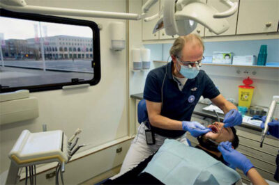
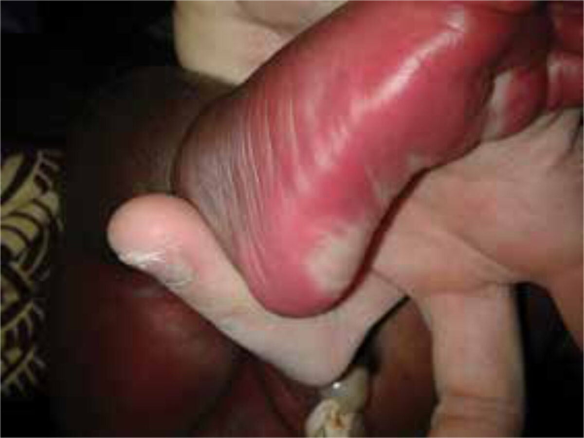
A child was born full-term in the delivery room of the Milange hospital. The pregnancy and birth were without complications and the mother already had two other healthy children. Immediately after birth, discolouration of the skin was observed. Throughout her whole body excluding the head, her skin was erythematous. This erythema was only mild on her chest, abdomen and back, but more severe on the right arm and leg. In general there was no sharp edge with normal skin. Moreover, a grey-blue discolouration was noted on the right leg and right side of her abdomen. All skin lesions were not raised above the level of the skin (figure 1). Furthermore, a remarkable finding was the raised capillary refill; the photo was taken more than two seconds after releasing pressure (figure 2). Also, the right leg was edematous. The erythema seemed painless and on further physical examination, nothing else abnormal was found.
Consult Online was asked for a differential diagnosis and advice in treatment.

Setting
Milange hospital is a district hospital located in the north of Mozambique on the border of Malawi. Milange district has around 700,000 residents. The hospital has a capacity of 98 clinical beds. It collaborates closely with the local health care centre, in which the outpatient clinic of the hospital is situated. The nearest referral hospital is at the other side of the border in Mulanje, Malawi.
Answer of the specialists
Two of the specialists responded within a few days. One mentioned the Klippel-Trenaunay-Weber syndrome, which includes an extensive port-wine stain with underlying venous and/ or lymphatic malformations involving an extremity. Because of the blue macula in addition to the capillary malformation, the diagnosis phacomatosis pigmentovascularis was said to be most likely. For the skin lesions, no treatment is available nor is it necessary. However, this condition is associated with, among others, neurologic, ocular and skeletal disorders. The advice of the specialist was to perform further examination on co-morbidities.
Background
Phacomatosis pigmentovascularis (PPV) is a rare, congenital disorder, characterized mainly by the presence of capillary malformations and pigmentary nevi. Since the first case, described in 1947, only around 245 other cases have been published [1,3]. Though the precise etiology remains unclear, it is thought to be based on a developmental abnormality of the vasomotor nerves and melanocytes, both derived from the neural crest [1]. Dermal melanocytosis occurs when fetal melanocytes fail to migrate to the dermis, causing a mongolian spot or blue nevus. The capillary malformation is present in most PPV types and is usually extensive. Furthermore, as many as half of the patients have other associated cutaneous lesions, most commonly nevus anemicus and café-au-lait spots [1].
Multiple types of PPV exist, but the most common is phacomatosis cesioflammea [1,2]. There are several classification systems. The first classification was developed by Hasegawa [2] and distinguishes type I-V. In addition, “a” or “b” are added to indicate extra-cutaneous involvement. Currently, the most practical classification is Happle’s system [1]. In this simplified, descriptive, classification system, there is no distinction between whether or not there are extra-cutaneous anomalies present (table 1).
It has been estimated that approximately 50 percent of patients have extra-cutaneous involvements, which are, as previously mentioned, mainly neurologic, ocular and skeletal disorders [1,2]. Neurologic abnormalities will usually be present in the first few months of life. These include among others, psychomotor retardation, seizures, intracranial calcifications, or cerebral atrophy. Common related ocular disorders are blue-gray scleral discolouration or glaucoma. There are several skeletal disorders described, such as discrepancy in the length of extremities, and scoliosis [3]. PPV without systemic involvement has a benign course and does not require treatment. The life expectancy depends on the presence and treatment of other associated conditions [1,3].
Table 1 Classification according to Happle [1]
| Type | Happle’s classification | Vascular lesion | Pigmentary lesion |
|---|---|---|---|
| I a/b | Non-existing** | Nevus flammeus (port-wine stain) | – |
| II a/b | Cesioflammea | Nevus flammeus (port-wine stain) and/or nevus anemicus | Mongolian spot |
| III a/b | Spilorosea | Nevus flammeus (port-wine stain) and/or nevus anemicus | Nevus spilus |
| IV a/b | unclassifiable type | Various types of vascular nevi | Mongolian spot, nevus spilus |
| V a/b | Cesiomarmorata | Cutis marmorata telangiectasia congenital | Mongolian spot |
Follow-up
At the time of discharge, the skin abnormalities seemed to improve slightly, as well as the edema of the right leg. The patient will be seen in the outpatient clinic for follow-up and is referred to a hospital in Malawi for further examination on co-morbidities. In conclusion, extensive skin abnormalities in a neonate should alert to conditions such as phacomatosis pigmentovascularis and one should be aware that extra-cutaneous involvement might be present.
References
- Phacomatosis pigmentovascularis revisited and reclassified. Rudolf R Happle, Archives of Dermatology 141(3) 2005 -3 0003-987X.
- Hasegawa Y, Yasuhara M. Phakomatosis pigmentovascularis type IVa. Arch Dermatol 1985; 121: 651-655.
- Fernández-Guarino M, Boixeda P, de Las Heras E, et al. Phakomatosis pigmentovascularis: Clinical findings in 15 patients and review of the literature. J Am Acad Dermatol 2008; 58:88.