


Main content
Setting
This case is set in a hospital run by Médecins sans Frontières, amidst an IDP (internally displaced people) camp in Bentiu, South Sudan. The camp is situated within the war zone and is inhabited by 130,000 people, some of whom have been on the run for months. The MSF hospital is their only source of medical help, providing basic facilities such as simple laboratory tests, ultrasound, and wards for a total of 100 inpatients. Among the Sudanese population, there is a high prevalence of malnutrition and tuberculosis. Visceral leishmaniasis is common among IDP from specific counties. Currently, there is an outbreak of malaria and hepatitis E. HIV prevalence is low, although the exact percentage among IDP is unknown.
Case
A 22-year old male patient presented at the outpatient clinic with a rash affecting the whole body. It was most prominent in the face, where the rash had started a few months earlier. It was not painful, itchy, or ulcerating, nor did it demonstrate purulent exudate. Apart from the rash, the patient was well. There was no fever. The patient had completed treatment for visceral leishmaniasis 8 months previously (sodium stibogluconate and paromomycin) and currently was not using any traditional medicine. His HIV status was unknown.
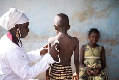
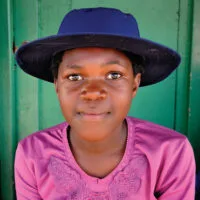
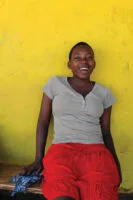
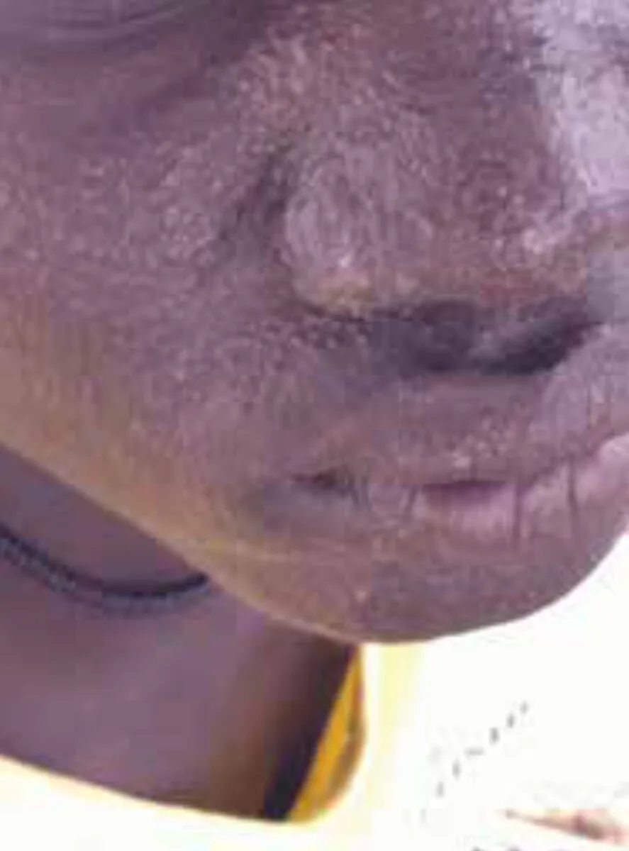
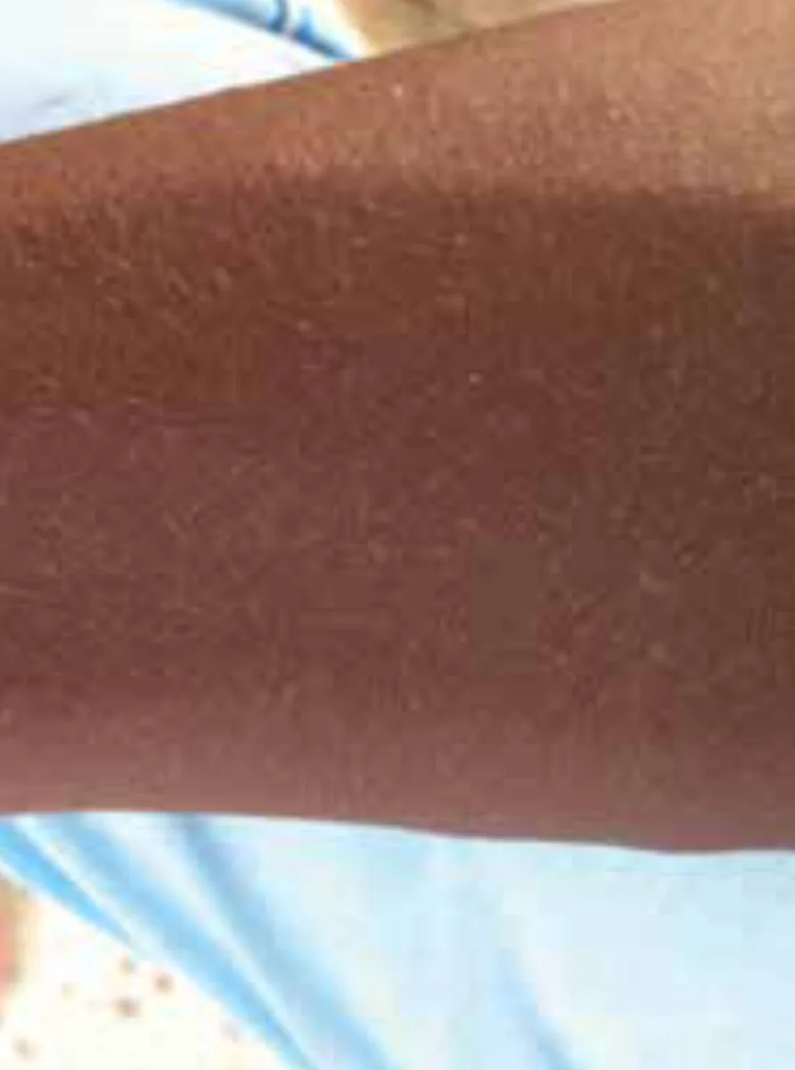
On physical examination, a papular rash was seen affecting the entire face, including the lips (Figure 1). Furthermore, there was bilateral conjunctivitis. On the chest and arms, small macules were found with less density than the facial rash (Figure 2). There was no lymphadenopathy or hepatosplenomegaly.
Specialist advice
The dermatologists were consulted for advice on the differential diagnosis as well as the treatment of this particular case. They suggested post-kala-azar dermal leishmaniasis (PKDL) as the most probable diagnosis and advised treatment with AmBisome or sodium stibogluconate, depending on local availability. As a differential diagnosis, the dermatologists proposed lepromatous leprosy or lichen nitidus.
Treatment
The patient was started on AmBisome. After 20 days, the rash had disappeared and the skin was near normal, much to the satisfaction of the patient.
Background
Visceral leishmaniasis (VL), in the Sudan known as kala-azar, presents with fever, weight loss and hepatosplenomegaly. It is caused by the parasite Leishmania, which is transmitted by the female sandfly. Post-kala-azar dermal leishmaniasis (PKDL) is a dermatological complication of VL, although it occasionally occurs without a previous history of VL.
Epidemiology and pathophysiology
PKDL occurs in East Africa and the Indian subcontinent, with marked differences in presentation [1]. In Sudan, it occurs in up to 50% of cases with VL. The disease-free interval between the successful treatment of VL and developing PKDL is 2 weeks up to 6 months. In India, PKDL is less common, affecting 5-10% of VL cases and presenting 2 to 3 years after VL.
PKDL is predominantly associated with VL caused by Leishmania donovani, although it may occur sporadically in Leishmania infantum [2,3]. The disease is thought to be immunologically mediated, showing a mixed Th2 (as found in VL) and Th1 (as found after treatment of VL) immune response [1,3].
Potential risk factors are young age (5-17 years), inadequate treatment of VL (short duration, low dose or mono therapy), comorbidity (including HIV), malnutrition, and exposure to UV light. [3] The severity of PKDL is determined by age and disease-free interval, with more severe disease occurring among young children after a shorter interval.
Clinical manifestations and diagnosis
PKDL is characterized by a skin rash without fever or other symptoms of systemic disease [3]. A combination of symmetrical papules, macules and nodules may be found. The lesions typically start in the face, may spread to the trunk and upper extremities, and may ultimately become generalized. In more severe cases, subsequent crusting, ulceration, and involvement of lips and palate may occur. In India, there may be an erythematous rash in the butterfly area of the face.
Other post-kala-azar manifestations include mucosal leishmaniasis, laryngitis, colitis, uveitis, conjunctivitis and blepharitis. In some patients, PKDL occurs during (treatment of) VL (para-kala-azar dermal leishmaniasis) [1].
PKDL can be diagnosed by identifying the parasites in slit-skin smears or biopsies [2,3]. Serological tests are of less clinical value, as they are likely to remain positive due to persisting antibodies after VL infection.
The differential diagnosis of PKDL is extensive and includes leprosy, pityriasis versicolor/alba, vitiligo, scabies, measles, acne vulgaris, lupus vulgaris, miliaria rubra, secondary syphilis and nutritional deficiencies [3,4].
Therapy
PKDL regresses spontaneously within a year in the majority (85%) of cases in Sudan. Therefore, patients are only treated in the case of severe disease, lesions lasting longer than 12 months, anterior uveitis or feeding difficulties (oral lesions in young children). In India, however, all patients are treated. It is thought that PKDL patients may act as reservoirs for leishmanial parasites and can therefore be a source of transmission; however the evidence for this is not conclusive.
Different treatment strategies have been proposed. First choice of treatment is AmBisome (liposomal amphotericin B, 2.5 mg/kg/day intravenously for 20 days), as it is associated with a high cure rate, short duration of therapy, and negligible side effects. Second choice is sodium stibogluconate (SSG, 20 mg/kg/day for 40 days), which has several toxicities and requires a prolonged hospital stay and painful injections. It may be combined with paromomycin. Because of the incomplete immunological response, immunochemotherapy has been studied with promising results. In Asia, miltefosine (150 mg/day for 60 days or 100 mg/day for 90 days) is the standard treatment; this has not been studied in Africa. Azoles, such as itraconazole or ketoconazole, were not found to be effective.
Post-kala-azar ophthalmological complications are often missed and may cause blindness in the case of uveitis. Steroid containing eye drops should be given in addition to antileishmanial therapy.
Outcome
The most important outcome of successful treatment is the disappearance of parasites from the skin. However, clinical response is the most important goal for the patient and is easier to monitor. Lesions disappear after 120 to 200 days (nodules/papules and macules, respectively), those around the mouth remaining longest [1]. In some cases, scars or depigmentation may remain. As in VL, patients will develop immunity for leishmaniasis after treatment, although rarely relapses of PKDL or VL may occur [1].
References
- Zijlstra EE, Musa AM, Khalil EAG et al. Post-kala-azar dermal leishmaniasis. Lancet Infectious Disease 2003;3:87-98.
- Ganguly S, Das NK, Barbhuiya JN et al. Post-kala-azar dermal leishmaniasis an overview. International Journal of Dermatology 2010;49:921-931.
- World Health Organization. Post-kala-azar dermal leishmaniasis: a manual for case management and control. Report of a WHO consultative meeting. India 2012.
- Zijlstra EE, Khalil EAG, Kager PA et al. Post-kala-azar dermal leishmaniasis in the Sudan: clinical presentation and differential diagnosis. British Journal of Dermatology 2000;143:136-143.
- Musa AM, Khalil EAG, Younis BM et al. Treatment-based strategy for the management of post-kala-azar dermal leishmaniasis patients in the Sudan. Journal of Tropical Medicine 2013; 2013:708391.