
Main content
Setting
Saint Theresa hospital is a district hospital in the Copperbelt, a region in the north of Zambia known for its copper mining. Patients are mostly farmers and the majority are poor. The nearest city is located 25 kms away. Annually, this hospital provides health care to 23,000 patients, and has a capacity of 100 beds. There is no X-ray facility available in the hospital; once a week patients are transported to another hospital for imaging.
Case report
A two-year-old boy complained of lower back pain after a fall. Because the X-ray of the lumbar spine showed no signs of a fracture, and no other abnormalities were seen during examination the parents were advised to let the child rest at home. After a week of watchful waiting the child returned to the physiotherapist, the pain had increased and he was not able to walk anymore. Moreover, the child had a fever and a cough. He was therefore sent to the hospital.
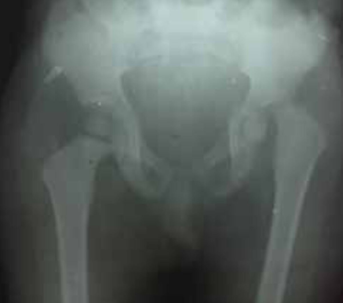
During physical examination he appeared to be low-weight for his age, pale and irritable; rhonchi were heard when auscultating the lungs. He was able to stand up, but unable to walk. The WBC was 9500/mm³. Because of the combination of fever, cough and abnormal breath sounds, empirical antibiotic treatment for pneumonia was started. However, a few days later the chest X-ray showed no abnormalities. Subsequently the child experienced pain in the left hip; he still could not walk. Therefore, it was suspected that the child could suffer from septic arthritis of the left hip and the antibiotics were changed into cloxacillin and ceftriaxone. After seven days of antibiotic treatment, the child remained painful and feverish. The WBC at that time was 2500/mm³. An X-ray of the left hip was done and it showed a complete epiphysiolysis of the femoral head (figure 1). Retrospectively, the left hip could also be seen on the lumbar spine X-ray taken on presentation. At that time the left hip joint showed no abnormalities.
Consult online was asked for advice on further treatment.

Advice from the specialists
Two specialists responded within a day. They both agreed that this was presumably a case of septic arthritis and they emphasized that this condition is difficult to diagnose in young children. In most cases, it is associated with osteomyelitis of the femoral head. Furthermore, the increased intra-articular pressure due to the infection, can compromise the blood supply to the femoral head, resulting in avascular necrosis and epiphysiolysis.
Their advice was to perform an arthrotomy as soon as possible. This enables decompression of the joint, debridement of the infected tissue and irrigation of the joint space. This should result in reduction of fever and by releasing the pressure, necrosis will hopefully be prevented. The specialists warned that necrosis might already have taken place. Their advice was also to place the child in traction and to continue antibiotic treatment.
Treatment and follow-up
The next day, arthrotomy was performed, with extensive irrigation of the joint. No intra-articular pus was found, but it was suggested that pus might already have turned into debris. A drain was left in the joint space. The child was placed in traction for 4 weeks and antibiotic treatment was continued. Pain and fever subsided. After a period of 4 weeks, a new X-ray unfortunately did not show much improvement in the position of the femoral head (figure 2). The child started to mobilize, which was again painful. Since there were no signs of infection anymore, and because of his overall good general condition, he was discharged from the hospital. Mother and child have never returned to the outpatient clinic, so unfortunately there is no information on the current condition of the child.
Discussion
When a child presents itself with acute pain in the lower limb area without a prior trauma, the main differential diagnosis includes transient synovitis (coxitis fugax), septic arthritis and Legg-Calvé-Perthes disease (1). Of all acute non-traumatic hip pathologies, transient synovitis (coxitis fugax) is the diagnosis with the highest incidence; however, differentiation between septic arthritis and transient synovitis of the hip in children is essential. Transient synovitis is self-limiting, whereas septic arthritis is treated with operative drainage and antibiotics, and can lead to osteonecrosis (and epiphysiolysis), growth arrest, and sepsis.
Both septic arthritis and transient synovitis present with pain during weight bearing as a cardinal feature. However, findings related to the involved joint may be subtle, especially in young children. In addition to pain other clinical features include fever, malaise, poor appetite, irritability and tachycardia and these can be present within the first few days of infection (1).
In order to distinguish septic arthritis from other non-traumatic hip pathologies, Kocher et al. (2,3) described four predicting factors for septic arthritis: a history of fever, non-weight-bearing, an erythrocyte sedimentation rate (ESR) of at least forty millimetres per hour, and a serum white blood-cell count of more than 12000 cells /mm³ (1). In this case, there was no ESR done, but the child did meet with two predicting factors, namely fever and non-weight-bearing. This means a predicting probability of 57.8%. In the advanced stage, the white blood cell count was raised to 25000 cells/mm³, which suggests a predicting probability of 95% (2,3).
Currently, the most common cause of septic arthritis of the hip joint in children is Staphylococcus aureus; however, in sub-Saharan countries, Salmonellae have a high prevalence (4). Children with HIV are more at risk, as well as children with sickle cell disease (4). The diagnosis septic arthritis can be confirmed with an ultrasound-guided aspiration of synovial fluid. This should be performed as soon as possible, when septic arthritis is suspected. Antibiotic treatment should start right after aspiration. If septic arthritis is highly suspected, for example as it was in this case, when there is already osteonecrosis seen on the X-ray, irrigation of the joint should be performed as soon as possible (1).
The functional prognosis of septic arthritis with osteonecrosis is poor. This child will presumably have a dislocated joint and severe limb shortening. In the above-mentioned case, we tried to prevent this with long-duration of traction, but unfortunately, without a satisfactory outcome. Eventually, a painless ankyloses may develop, which would probably be the best possible scenario. There are no therapeutic options in children, and the final situation should be awaited for at least ten years (1,4).
In conclusion, septic arthritis does not run a typical course in young children, which makes it a disease difficult to diagnose with potentially major complications, especially in a low-resource setting.
References
- Rutz E, Spoerri M. Septic arthritis of the paediatric hip – A review of current diagnostic approaches and therapeutic concepts. Acta Orthop. Belg 2013; 79: 123-134.
- Mininder S, Kocher MS, Zurokowski D, Kasser JR. Differentiating Between Septic Arthritis and Transient Synovitis of the Hip in Children: An Evidence-Based Clinical Prediction Algorithm. J Bone Joint Surg Am 1999;81:1662-70.
- Mininder S; Kocher MS. Validation of a clinical prediction rule for the differentiation between septic arthritis and transient synovitis of the hip in children. J Bone Joint Sur 2004;8:86-A (8):1629-35
- Christopher BD, Lavy CB. Septic arthritis in Western and sub-Saharan African children – a review. Internat Orthopaed 2007;4:31(2):137-44