Main content
The clubfoot is most often a congenital deformity that occurs in 1 to 2 of ev-ery 1,000 neonates. Ideally, treatment of the clubfoot is started at birth, as is done in the modern world. However, with the limited finances and access to medical care and /or hospital resources in Low- and Middle-Income Countries (LMICs), the majority of the clubfoot patients in the LMICs are inadequately treated or neglected.
Neglected clubfoot can have a dramatic effect on the quality of life. Physical impairment results in decreased ambulation and disability will lead to dependency in ac-tivities of daily living with significant economic impact on both the family and the village. Furthermore, the visible physical and functional differences in individuals with clubfoot are associated with considerable social stigma. Children with locomotor disability are less likely to be able to access education and a proper job. Girls are even more vulnerable to social, physical and sexual abuse, and are less likely to achieve education than boys with the disability.
The deformity affects boys twice as much as girls, and twice as many cases are bilateral.
Pathology
The pathology affects especially the soft tissue around the posterior and medial site of the hindfoot. There is always stiff-ness, but some feet are stiffer than others. The deformity of the clubfoot when not treated is progressively aggravating over lifetime.
The deformity consists of an Equinus and Varus position in the hindfoot and Cavus -Adductus deformity in the midfoot.
Diagnosis
Early recognition of the deformity and correct diagnosis is of the greatest importance: congenital or acquired clubfoot. A classification and scoring of the foot is done and treatment has to start as soon as possible.
The primary treatment
The primary treatment of the clubfoot is nonsurgical and consists of specific manipulations of the foot followed by a specific method of casting during 5 to 6 weeks in progressive correction as described by Ponseti. This casting method needs training of the local paramedic staff.
If insufficient dorsiflexion of the foot is obtained after 5 to 6 weeks of progressive corrective casting a percutaneous tenot-omy of the Achilles tendon is necessary, followed by a further minimum of 3 weeks of casting.
The casting period has to be followed by a specific method of bracing for up to 3 years to avoid recurrences. If relapses occur within the first 3 years it is mainly due to the brace not having been worn !! Also local orthopedic technicians need to be trained to make and be able to adapt these braces while the child is growing rapidly.
As a non-invasive technique for the management of congenital clubfoot, the Ponseti method has been proven to be a superior method of correcting clubfoot deformity and avoids major surgical intervention. With this method, a long-term study by Cooper and Diez reported 78% good and excellent results at a minimum follow-up of 25 years.
The treatment of the neglected clubfoot
The treatment of the neglected clubfoot is surgical, to a high percentage, and becomes more difficult the older the patient is. The results of the surgery highly depend on the age of the patient and the stiffness of the non-treated or undertreated foot (recurrence).
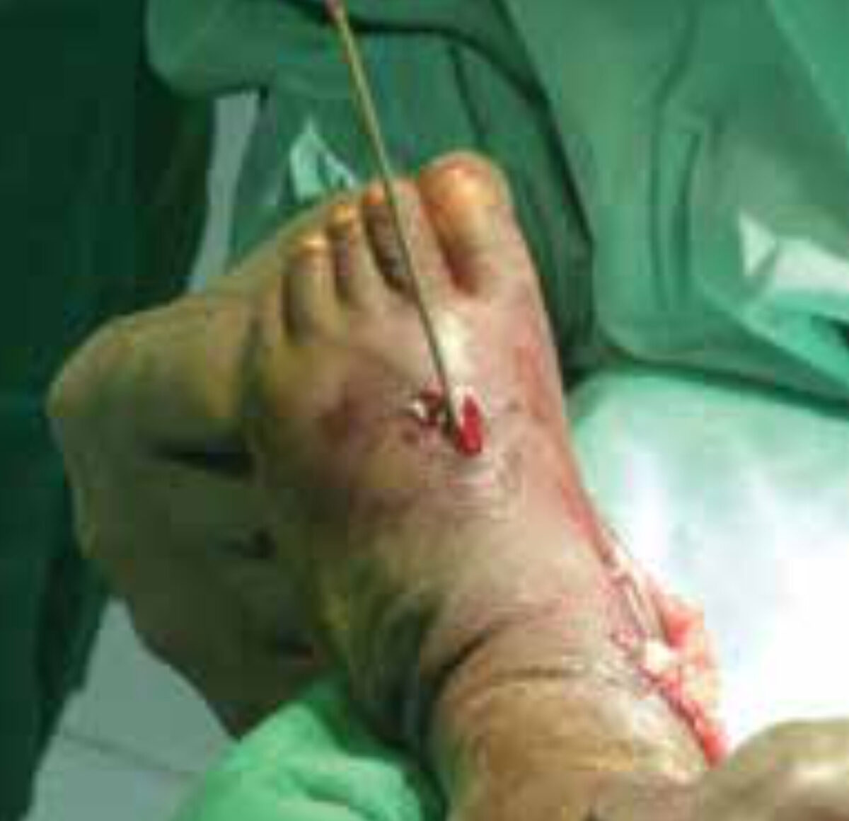
Under the age of 5 years the surgical procedure consists mostly of a release of the posterior medial soft tissues and capsular structures. The reduction has be fully and is best hold with a K-wire (figure 4). Immediate postoperative POP casts during 6 weeks are given, followed by bracing of the feet and lower limb in 10°of dorsiflexion, external rotation and abduction overnight and up to 3 years.
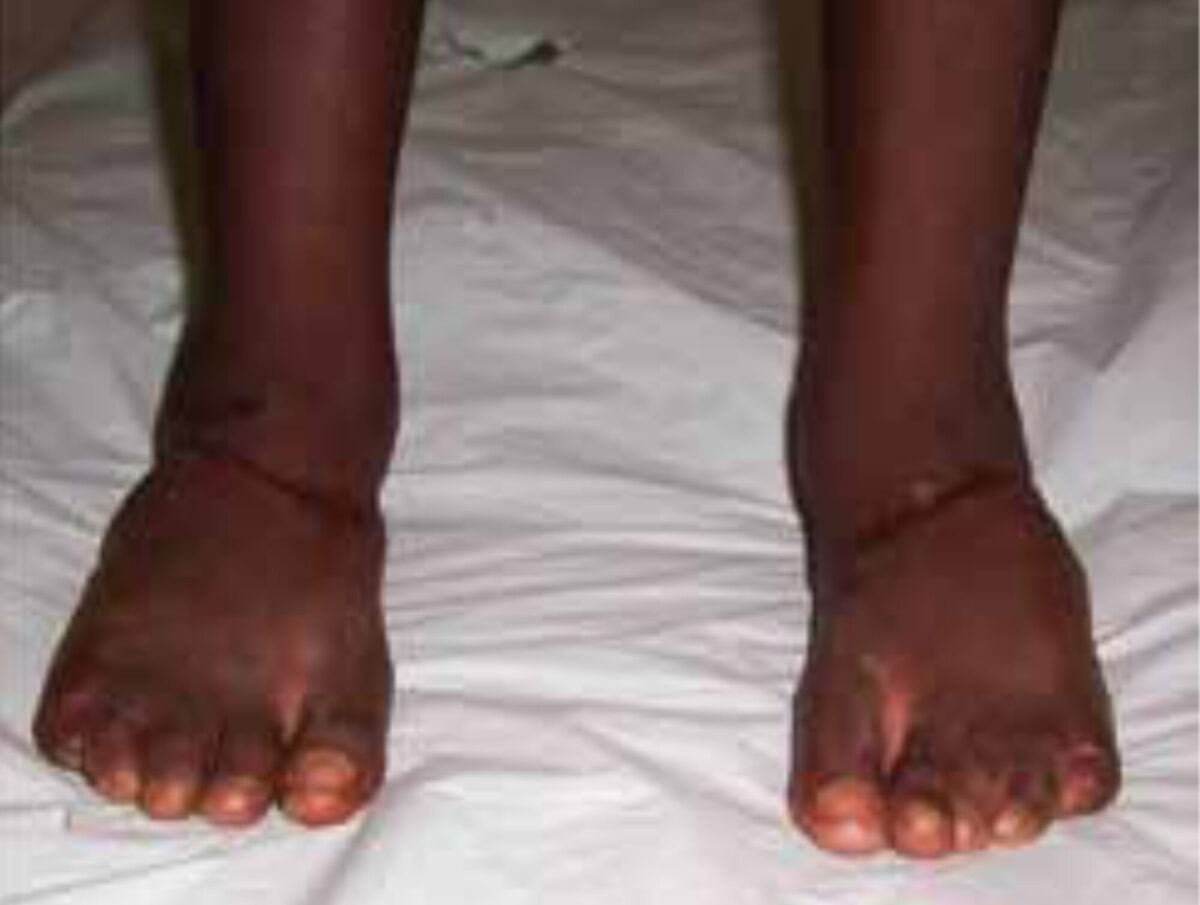
Seldom bony procedures are necessary before the age of 5. In age group 5 to 10 years soft tissue procedures may need to be supplemented by osteotomies of the calcaneum, navicular or cuboid bones (figure 2).
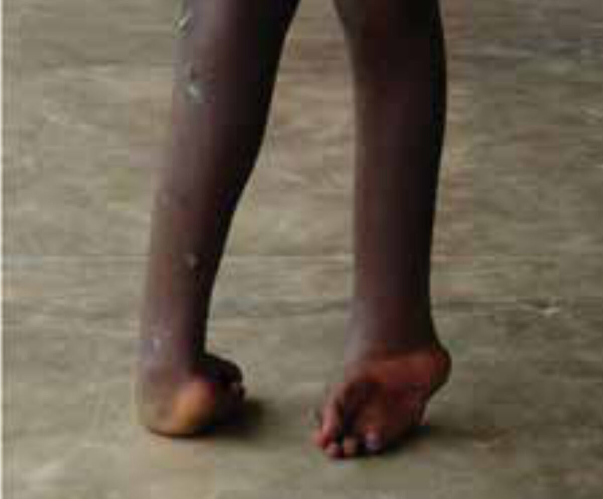
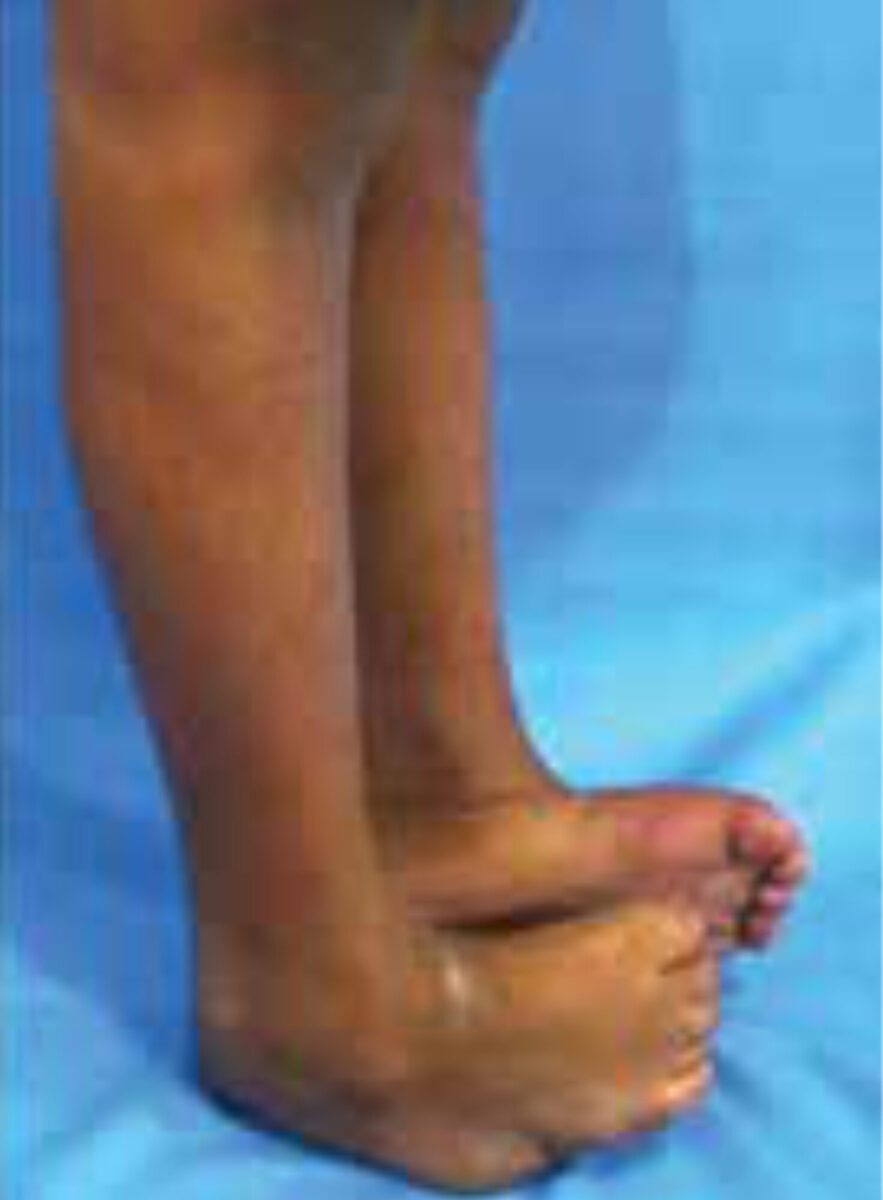
In adolescence surgery will include soft tissue release as well as bony procedures. A triple arthrodesis combined with Achilles lengthening, rotational skin flap will often offer the best pos-sibility of tridimensional correction to obtain a plantigrade foot (figure 1 and 3).
In the very severe adult neglected clubfoot, even a triple arthrodesis will not be able to give enough correction, due to vascular impairment and a talectomy or tibio-calcaneal fusion may then provide an adequate solution.
A skin rotation flap needs to be considered at any time to improve the reduction of the foot when the medial skin is with-holding, as it will also help the wound closure.
Postoperative casting is very important until full correction and healing of the bony procedure are obtained. Maintaining the correction with a brace and special footwear is just as impor-tant as the surgery.
In the paralytic feet tendon transfers should not be forgotten, and will be added to the above described procedures.
Conclusion
A clubfoot has to be recognized as soon as possible and treat-ment should ideally be started at birth by conservative meth-ods. The Ponseti management is the first choice treatment, in the developed as well as in the developing countries.
In childhood treatment concerns mainly soft tissue releases. In adolescence surgery is the better option, with triple arthrodesis being one of the important procedures to obtain a plantigrade foot.
These surgical procedures are difficult and need skilled surgeons and are not free from complications!!! Education of trained paramedical people and bedside training of doctors in the surgical procedures together with specific extended postop-erative care, will help to get the same good results in the care of the clubfoot as in the developed countries where the deformity hardly exists after the age of one year.
Further reading
- Clubfoot: Ponseti Management, 2009, third edition, available free from www.global-help.org
- Pirani S, Naddumba E, Mathias R et al. Towards effective Ponseti clubfoot care-The Uganda sustainable clubfoot care project: Clin Orthop Relat Res 2009;467:1154-63.
- Ponseti IV, Treatment of congenital club-foot. J. Bone Joint Surg. 1992;74(3):448-54.
- Ponseti IV, Congenital clubfoot. Funda-mentals of treatment, New York Oxford University Press 1996, available free from www.ponsiti.info
- Ponseti IV, Clubfoot Management, J Pediatr Orthop 2000;20(6):699-700.
- Cooper DM, Dietz FR, Treatment of idio-pathic clubfoot: a thirty year follow-up note. J Bone Joint Surg Am 1995;77:1477-89.
- Turco VJ, Resistant congenital clubfoot: one stage posteromedial release with internal fixation. A follow-up report of fifteen years experience. J Bone Joint Surg Am 1979;61:805-14.
- Baxter Willis R, Al-Hunaishel M, Guerra L, et al. What proportion of patients need extensive surgery after failure of the Ponseti technique for clubfoot? Clin Orthop Relat Res 2009;467:1294-97.
- Bensahel H, Csukonyi Z, Desgrippes Y, et al. Surgery in residual clubfoot: one-stage medio posterior release ‘a la carte.’ J Pediatr Orthop 1987;7:145-8.
- Pirani S, Zeznik L, Hodges D, Magnetic resonance imaging study of the congenital clubfoot treated with the Ponseti method.J Pediatr Orthop 2001;21:719-26.
- Dyer PJ, Davis N, The role of the Pirani scoring system in the management of club foot by the Ponseti method. J Bone Joint Surg Br 2006;88:1082-4.
- Mittal RL, The surgical management of resistant club foot by rotation skin flap and extensive soft tissue release. Int. Orthop 1987;11(3);189-92.