


Main content
Lupus erythematosus (LE) is an autoimmune disease with very diverse clinical manifestations, potentially affecting major internal organs such as kidney and heart, but more commonly the joints and the skin. The disease has a worldwide distribution but certain populations like Afro-Americans in the USA and people of Afro-Caribbean descent have a higher risk of developing systemic lupus erythematosus (SLE) compared with other groups. These populations may also have an earlier onset and more serious disease manifestations of SLE. This is probably due to the genetic predisposition for LE in these patients. SLE is more often seen in women; the male: female ratio is 1:9.
Pathogenesis of LE
There is an aberrant immune response to cellular and nuclear self-antigens that may be triggered by environmental factors in a genetically predisposed individual. The various tissues involved show chronic inflammation that eventually may lead to organ damage and loss of function.
Apoptosis is a strictly regulated process of programmed cell death that follows characteristic biochemical and morphological features. This type of cell death is contrary to necrotic cell death. It is a process that is vital for tissue homeostasis and apoptotic cellular remains are continuously cleared in a non-inflammatory way by macrophages or dendritic cells. This removal of apoptotic debris is associated with the induction of tolerance to the constituents of the material. In patients with LE there are indications that there is an increased amount of apoptotic material and that tolerance for self-antigens is being lost. Apoptotic cellular antigens accumulate in the body and are processed in an inflammatory way with activation of T- and B cells, production of autoantibodies and the deposition of antigen-antibody complex in tissues.
An important environmental trigger is ultraviolet radiation (UVR), which can induce apoptosis of keratinocytes in the skin within a few hours after UV exposure.
Pro-inflammatory clearance of apoptotic keratinocytes may be an important reason for UVR-induced skin lesions in patients with LE. Studies have found that more than 90% of patients with LE show some degree of photosensitivity. Therefore, it is important that all patients with LE are advised on photoprotection. Sunprotection may lead to vitamin D deficiency. Studies show an inverse correlation between SLE disease activity and serum vitamin D concentration although the effect of increasing vitamin D levels on disease activity is in general rather small.
There are many drugs that may trigger or worsen manifestations of LE and it is striking that the offending drug may have been used for several months or sometimes years. (Table 1) Patients with drug-induced LE often have more extensive skin symptoms than those with the idiopathic form and these lesions may persist for weeks or months after stopping the offending drug. These patients also frequently complain of malaise, arthralgia or fever; however, the central nervous system or the kidneys are not involved.
Table 1. Drugs that may induce lupus erythematosus
- Calcium-antagonists
- Thiazide diuretics
- ACE inhibitors
- Beta blockers
- Terbinafin
- TNF-alpha blockers
- Protonpump inhibitors
- Statines
- Antibiotics
The diagnosis of cutaneous LE is confirmed by histopathological examination of a skin biopsy of a suspected skin lesion. This will often show an interface dermatitis with necrotic keratinocytes and a lymphocytic perivascular infiltrate and gradual thinning of the epidermis. It is not helpful to determine the ANA titre because also in the general population positive titres can be found.
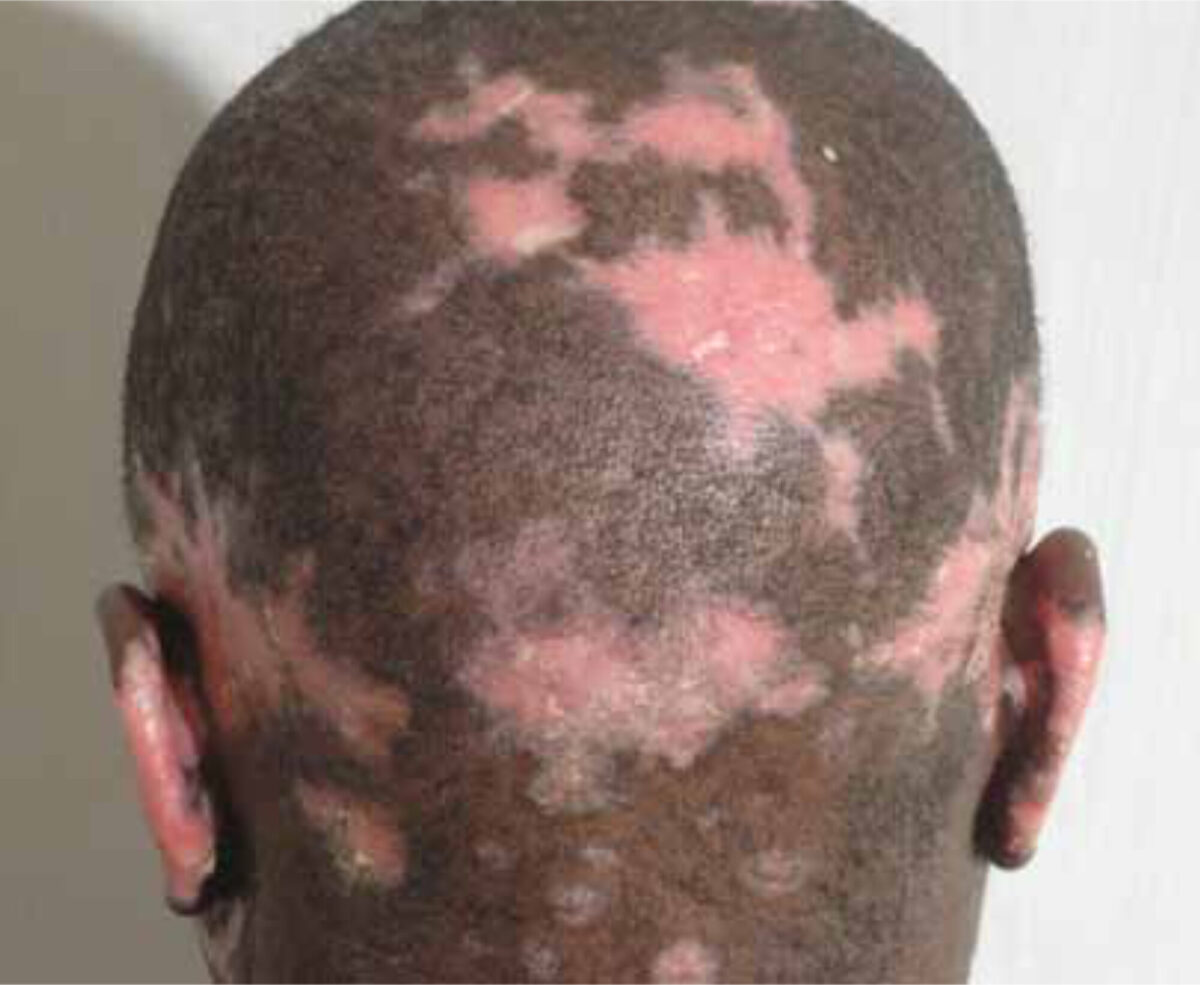
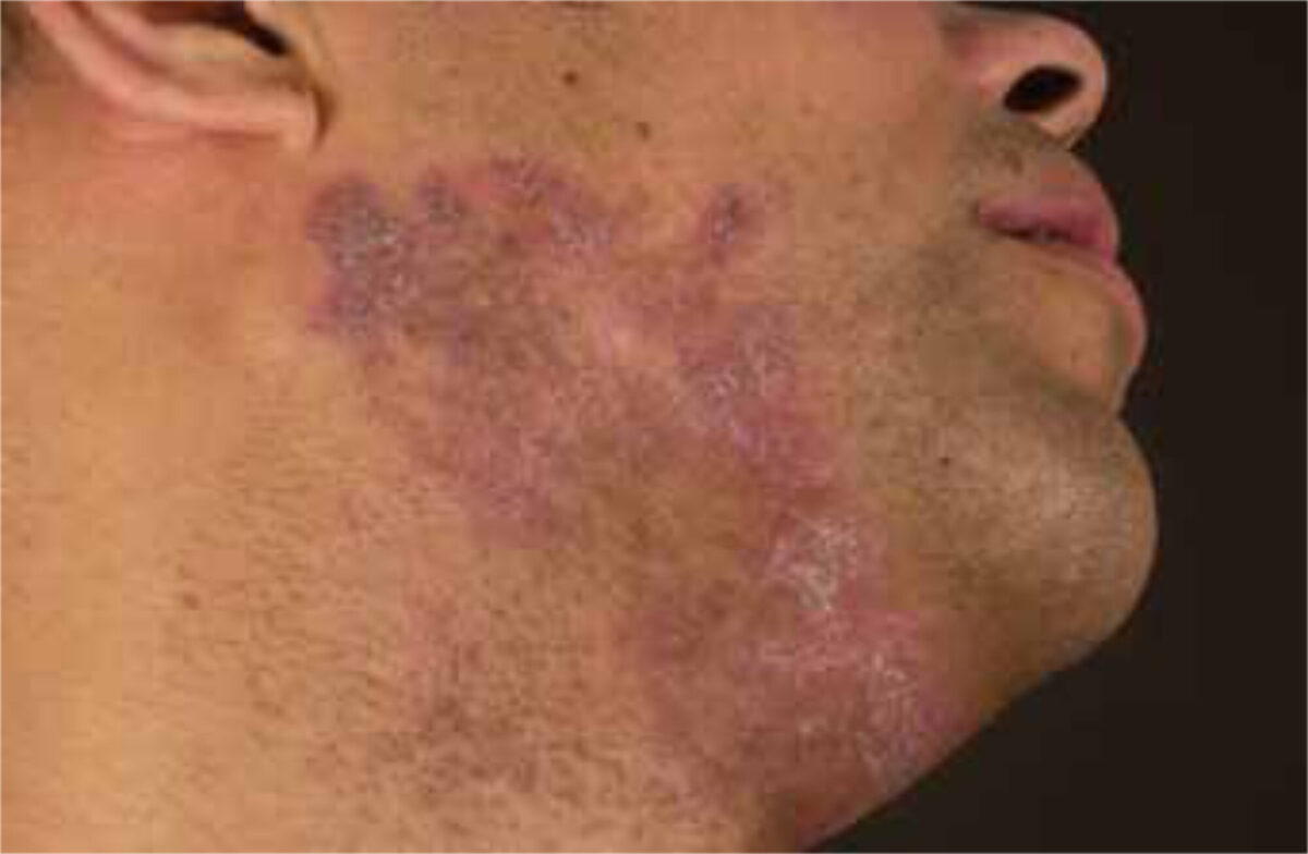
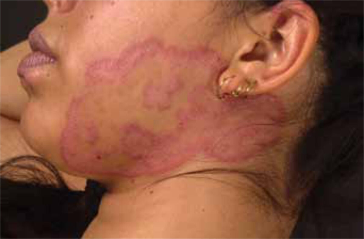
Cutaneous LE is often classified as acute, subacute and chronic cutaneous LE and other subtypes such as chilblain lupus, tumid lupus and neonatal lupus. Non-specific manifestations of cutaneous LE include vasculitis, periungual telangiectasia, Raynaud’s phenomenon, non-scarring alopecia and others.
Acute cutaneous LE shows erythematous papules and plaques in face and neck area with erosions. The subacute form has noticeable erythematous scaling patches and plaques that are sometimes annular and are found especially on the trunk and the extensor surface of the upper extremities. Chronic cutaneous LE is manifested by circumscribed papules and plaques with hyperkeratosis and sometimes erosions, often in the face but sometimes more widespread over the body. These lesions heal with scar formation. It is not always feasible to establish one specific diagnosis and there may be several subtypes present simultaneously. In these cases it is better to simply use cutaneous LE as a diagnosis. The risk of progression of cutaneous LE to SLE is about 5-15%. It is important to note that 17% of the patients with subacute cutaneous lupus erythematosus (SCLE) already have four criteria as suggested by the American College of Rheumatology for SLE.
Prevention
Patients with SLE are at an increased risk of cardiovascular problems and thrombo-embolic events. Smoking again increases these risks and also causes increased disease activity. Therefore, advice and counselling on how to stop smoking is very important. All patients with LE should receive advice on sun protection, such as behaviour or lifestyle change, protective clothing, and the application of sunscreens. When assessing a patient it is paramount to scrutinize the medications that are being used and stop drugs that potentially induce or exacerbate LE symptoms.
Treatment of LE
The goal of therapy in patients with LE in general is to reduce inflammation in all affected tissues or organs and furthermore to prevent damage that may evolve from these processes. The mainstays of treatment of cutaneous LE are topical corticosteroids and immune response modifiers, such as tacrolimus and pimecrolimus. Often potent corticosteroids such as betamethasone valerate are needed in order to reduce inflammation and if this alone is insufficient e.g. tacrolimus may be added. Hyperkeratotic forms of cutaneous LE may be treated with retinoids. If the inflammation is rather deep in the dermis or topical steroids are not effective systemic steroids may be necessary to control inflammation, preferably for short periods of time. Immunosuppressive drugs, such as methotrexate, azathioprine or mycophenolate mofetil, may be used if systemic steroids are contra-indicated or added to the therapeutic regimen as steroid-sparing agents. (Table 2)
Antimalarials and LE
Antimalarial treatment is gaining more prominence as a therapeutic tool and also as a potential measure to prevent progression to SLE. There is also increasing evidence that they reduce disease activity and mortality in patients with SLE. Therefore, all patients with SLE should use an antimalarial, preferably hydroxychloroquine, for prolonged periods of time, irrespective of disease activity, other medication used, and also during pregnancy.
Hydroxychloroquine is often given as a daily dose – 6.5 mg/kg lean body weight and chloroquine 3 mg/kg lean body weight. They are probably equally effective but chloroquine has more side-effects. A good way to commence with hydroxychloroquine is to start with 200mg daily and, if no side-effects occur, to increase the daily dose to its maximum level, also taking the kidney function into account. Opthalmological examination is warranted with continued chronic use and high cumulative doses because of the risk of retinopathy.
Table 2. Therapy of lupus erythematosus:
- Topical and intralesional corticosteroids
- Topical tacrolimus or pimecrolimus
- Antimalarials (hydroxychloroquine, chloroquine)
- Systemic corticosteroids
- Retinoids (Neotigason, Roaccutane)
- Immunosuppressive agents (methotrexate, mycophenolate mofetil, azathioprine)
- Dapson, thalidomide (lenalidomide), clofazimine
- Pulsed dye laser therapy
- Fumaric acid
- UVA-1, UV hardening therapy
- Biologicals and anti-cytokine therapy (rituximab, belimumab)
- Cyclophosphamide
- Intravenous immunoglobulin
- Stemcell transplantation
