
Main content
Setting
This case is from Haydom Lutheran Hospital, located in the village Haydom, Manyara region, Tanzania. The hospital is a regional referral center, and serves people from a catchment area of 2 million people. It has approximately 400 beds with capacity to conduct basic laboratory tests, cultures, X-rays, CT scans and ultrasounds. However, patients often do not have insurance and/or the financial means to pay for all required diagnostics and treatment, and choices need to be made. Several specialists in the hospital are available for consultations, including a urologist, gynaecologist, radiologist, paediatrician, internal medicine physician and three surgeons.
Case
A 35-year-old pregnant woman, gravida 5 para 4 living 4, presented in our setting with sudden onset of lower abdominal pain for one day. The pain was located in the periumbilical region and non-radiating. She also complained of hard stools since three days. The pain was not associated with other complaints and there was no history of trauma. She reported normal foetal movement, no per vaginal blood loss or leaking. She did not remember her last menstrual period and did not bring her antenatal clinic card. She had no previous medical or surgical history.
On examination, she was pale and her vital signs were normal (BP 129/74, PR 88, RR 26, SpO2 96%, RBG 8.4). Abdominal examination showed a fundal height of approximately 30 weeks gestational age, a soft uterus with tenderness around the umbilicus. The remaining systems were normal. Obstetric ultrasound showed a viable singleton pregnancy of 32 weeks gestational age in breech presentation with normal amniotic fluid and placenta. Laboratory findings showed a haemoglobin of 7.6 gr/dL, and elevated white blood cell count of 14.2 x 10⁹ while urinalysis was normal. The other laboratory results were unremarkable.
The patient was admitted in the obstetric department with clinical diagnosis of partial intestinal obstruction and conservative management was started. Corticosteroids injections were started for foetal lung maturation.
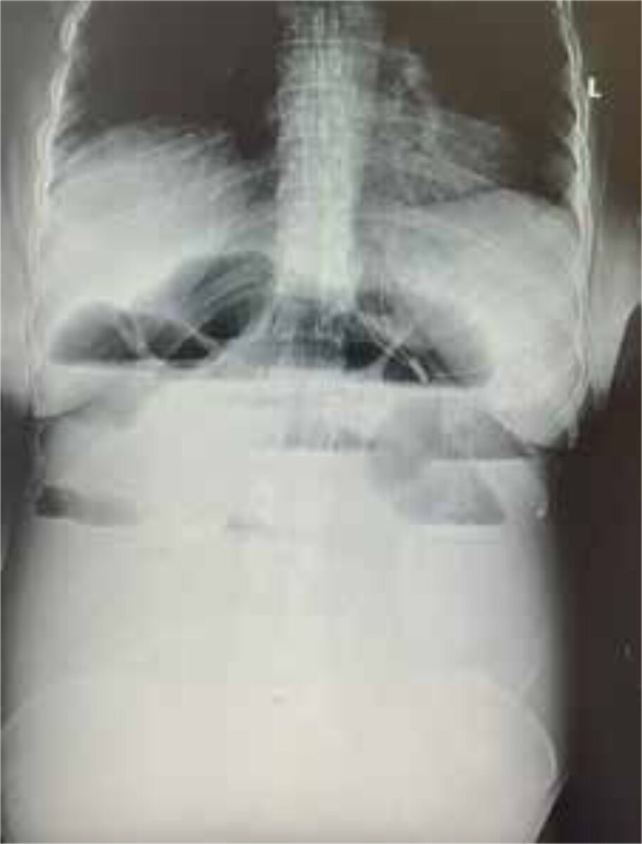
Two days after admission, the patient’s condition worsened due to increasing abdominal pain and vomiting of bilious materials with no passing of stools. On examination, she had normal vitals and foetal heart rate, but the abdomen was distended with tenderness around the umbilicus and epigastrium and exaggerated bowel sounds. Per vaginal examination showed a closed cervix and digital rectal examination an empty rectum. Additional laboratory findings showed negative MRDT and normal electrolytes, RBG, renal function and liver enzymes. Abdominal ultrasound showed multiple bowel loop dilatation suggestive of intestinal obstruction. There was no free fluid or other abnormalities detected. An abdominal X-ray was performed and showed multiple air fluid levels with bowel distention and no free air (Figure 1). Our provisional diagnosis was intestinal obstruction secondary to a sigmoid volvulus or adhesions, and conservative ileus management was started with nil per mouth, high volume iv fluids, nasogastric tube (NGT), catheter, serial enema, pain medication and broad spectrum antibiotics (iv ceftriaxone and iv metronidazole). Two blood units were prepared for transfusion.
After a short period of improving condition, four days after admission, the patient deteriorated despite conservative management, with increasing abdominal pain, generalised tenderness, distention and NGT draining faecal material. Following a discussion involving general surgeons and the obstetrician, the decision was reached for emergency exploratory laparotomy with caesarean section.
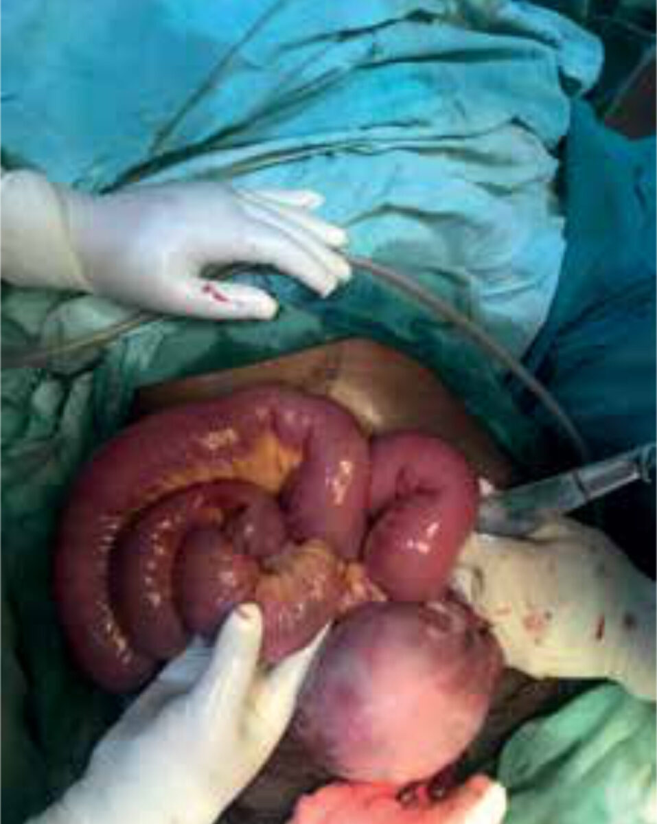
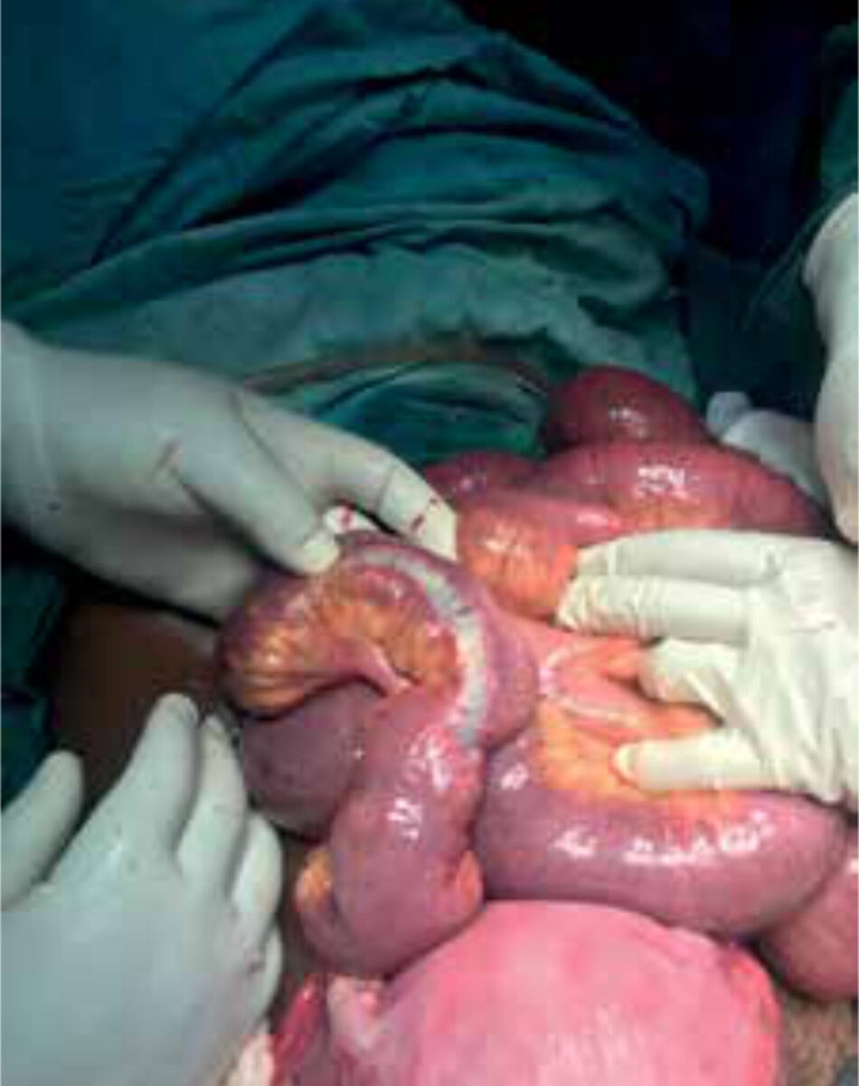
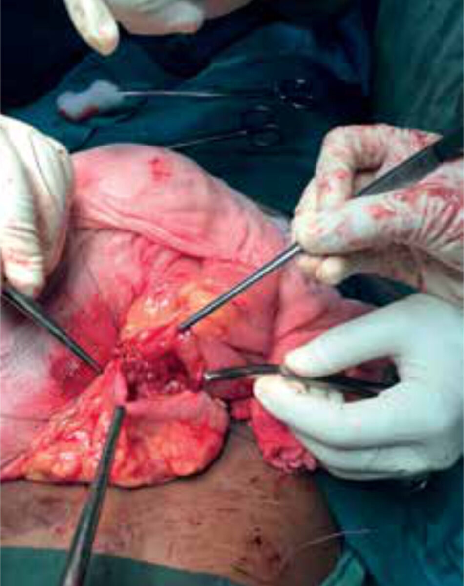
An extended midline incision was done and clear ascites was found. A caesarean section was performed. The neonate, weighing 1860 grams with Apgar scores of 4 in the 1st minute and 6 in the 5th minute, was resuscitated and transferred to NICU for further management. Furthermore, distended small bowels were found with an ileo-ileal intussusception (Figure 2) at 20 cm proximal from the ileocecal junction. An intraluminal white-ish non mobile firm tumour of approximately 2 cm, 40 cm proximal from the ileocecal junction, was found as the lead point (a lesion or variation in the intestine that is trapped by peristalsis and dragged into a distal segment of the intestine). There were no signs of intra-abdominal metastases. Then the ileo-ileal intussusception was reduced (Figure 3) and a resection of approximately 45 cm of ileum was done. This was followed by an end to side primary anastomosis of ileum with transverse colon (Figure 4). The ileum stump at the ileocecal junction was closed in two layers and the window between the ileo-loop and transverse colon was closed to prevent internal herniation. The abdominal cavity was washed and a drain was placed before closing the abdominal wall. Postoperatively her clinical condition improved quickly, and she was discharged the 4th postoperative day. The neonate was discharged ten days post-partum in good clinical condition.
Follow-up
One week after discharge, the patient came back to the OPD for follow-up and suture removal. She was in improving clinical condition, and repeated abdominal ultrasound was normal. The histopathology result of the biopsy revealed benign gastrointestinal stromal tumour (GIST).
Background
Intestinal intussusception is rare in adults and accounts for 1 to 5 percent of mechanical bowel obstructions. In pregnancy this condition is less common and only described in case reports. [1-7] Most cases occur during the third trimester. It is typically caused by a pathological lead point (lesion or variation) within the bowel, which is pulled forward by normal peristalsis, telescoping the affected segment of bowel into the lumen of the distal bowel segment. Patients with disorders associated with higher occurrence of infections and neoplastic conditions, e.g. HIV, have an increased incidence of intussusception, secondary to conditions such as lymphoid hyperplasia, Kaposi sarcoma and non-Hodgkin lymphoma that can serve as lead points. [8] The intussusception leads to venous and lymphatic congestion, causing oedema and eventually intestinal ischemia. [6] Complications can be life-threatening in the case of intestinal necrosis and perforation, thus requiring early diagnosis.
Intestinal obstruction in pregnancy is a surgical emergency which requires acute intervention. [9] It is associated with high incidence of foetal and maternal morbidity and mortality, with maternal and perinatal mortality of 6% and 26%, respectively. [2,3]
Classification
Intussusception in adults can be classified by aetiology[4,8]:
- Benign lesion (e.g. polyps, lymph nodes, lipoma, Meckel’s diverticulum, inflammatory bowel disease, adhesions or trauma)
- Malignant lesion (25%; Metastatic disease or primary small bowel tumour such as lymphoma, leiomyosarcoma, neuroendocrine tumour)
- Idiopathic (8-20%)
It can also be classified by location:
- Entero-enteric, which is limited to the small bowel
- Ileo-colic with prolapse of the terminal ileum into the ascending colon
- Colo-colic, which is limited to the large bowel. [4,8]
In this case, intestinal obstruction was caused by a benign gastrointestinal stromal tumour (GIST) in the ileum leading to ileo-ileal intussusception. GISTs are rare, typically benign, mesenchymal neoplasms of the gastrointestinal tract. [10] About 10 to 30% of GISTS progress to malignancy. [10] The dynamic of growth is exophytic, having the potential to invade the adjacent organs, and in some cases causing perforation into the peritoneal cavity. GISTs rarely cause intussusception or obstruction. [11]
Diagnosis
Adults often present with intermittent abdominal pain and symptoms of (partial) bowel obstruction such as nausea, bloating, vomiting, constipation and weight loss. These symptoms can be subtle and are non-specific and are encountered frequently during a normal pregnancy, making diagnosis potentially difficult. Moreover, the displacement of the bowel by the gravid uterus impedes the physical examination. [3] In the diagnostic and therapeutic process, the risks and benefits for both mother and foetus must be considered. [4] However, a delay in diagnosis of surgical abdomen in pregnant women leads to higher complication rates.[9] This is significantly more risky to the foetus than the radiation exposure, since ionising radiation exposures up to 0.05 Gy are usually safe in pregnancy. [12]
Plain abdominal and chest radiography in supine position could show the typical features of distal small bowel obstruction, like distended bowel loops. [3] Air under the diaphragm in erect position indicates visceral perforation. However, an abdominal CT scan is the most sensitive imaging modality in the diagnosis of adult intussusceptions, showing the location, cause and potential complications of bowel obstruction, like ischaemia or perforation. The distended loop of bowel appears thickened because it represents two layers of bowel. A “target sign” may be seen on the sagittal view of the abdominal CT, while on axial or coronal view, the intussusception will appear as a sausage-shaped soft tissue mass. [3,8] The lead point can be identified as well.
Abdominal ultrasonography may be useful for the diagnosis of small bowel obstruction in pregnant patients, since it is fast and avoids radiation, and in settings with limited resources. Ultrasound is limited by obesity and by poor visualisation of gas-filled structures, but it is more sensitive and specific than plain films for the diagnosis of small bowel obstruction. The sonographic findings of intussusception are similar to CT scan, showing a ‘target’ or ‘doughnut sign’ on transverse section, which consists of single or double anechoic rings surrounded by a central echogenic focus [2,5,6], and the pseudokidney or sandwich sign in the longitudinal view. [11]
If available, abdominal magnetic resonance imaging (MRI) is also an option for the assessment of small bowel obstruction in pregnant women and in children. However, more time is required for high-quality image acquisition and for repeated breath-holds, which limits the general applicability of MRI in patients with acute small bowel obstruction. [8]
In low-resource settings with limited access to imaging modalities, clinical history and physical exams with frequent re-evaluation should be the basis of early diagnosis and surgical management.[11,13]
Differential diagnosis intestinal obstruction in pregnancy
Intestinal obstruction in pregnancy has an incidence of between 1:2500 and 1:3500 deliveries, mostly during third trimester. It is most commonly caused by adhesions. [3,14] Other causes of intestinal obstruction in pregnancy include volvulus, intussusception, carcinoma, herniation, and acute appendicitis.[1,2] While adhesions may be the most common cause of intestinal obstruction in patients that have had previous abdominal surgeries, the possibility of other causes of intestinal obstruction must always be considered.
Treatment
In intussusception in pregnancy, timely diagnosis can prevent bowel ischaemia and reduce maternal morbidity and mortality. The combined expertise of the obstetrician, radiologist, and surgeon are needed to manage the pregnant patient. [2] In contrast to adhesional obstruction, where patients may be managed conservatively initially, for other causes of intestinal obstruction, including intussusception, surgery remains the definite management and an exact diagnosis can be made during the operation. [3,11,14] Surgical resection of the lead point and necrotic bowel is almost always required in intestinal obstruction during pregnancy. [2] Intra operative reduction can be attempted in small bowel intussusceptions provided that the segment involved is viable and a malignancy is not suspected. If intestinal obstruction occurs during the third trimester of pregnancy, a concomitant caesarean section should be considered to increase the chance of foetal survival and also because of the fundal height necessitating a caesarean section before bowel exploration. (9,12,15] Providing corticosteroids for foetal lung maturation can be considered if the gestational age is below 34 weeks, but should not cause a delay in surgical intervention. If no caesarean section is performed during laparotomy, the overall risks of premature labour are significant. [14]
Conclusion
Intussusception in pregnancy is a rare condition and can be a diagnostic challenge. Patients often present with very non-specific symptoms and unreliable physical exam findings. High index of suspicion and systematic re-evaluation of the pregnant patient with intestinal obstruction are essential for timely diagnosis and treatment, which is key for preventing complications and poor outcomes for mother and foetus. Surgery remains the definite management in adult intussusception, and an exact diagnosis can be made during the operation.
Contact: c/o MTredactie@nvtg.org
References
- Chang Y.T., Huang Y.S., Chan H.M., Huang C.J., Hsieh J.S., Huang T.J. Intestinal obstruction during pregnancy. Kaohsiung J. Med. Sci. 2006;22:20-23.
- Achour R, Harabi S, Neji K. Spontaneous acute intussusception in a pregnant woman. Case Rep Womens Health. 2016;13:6-8.
- Firdaus Hayati, Asyraf Mohd Zuki, et al. A peculiar case of intussusception in a pregnant woman: A diagnostic challenge. Radiology Case Reports. 2023;18(8): 2836-2839.
- Vaynshtein J, Guetta O, Replyansky I. Abdominal Pain in Pregnancy. JAMA Surg. 2019;154(2):176-177.
- Alexander D. Combes MBBS, Alexandra M. Limmer MBBS (Hons), Kurt Verschuer FRACS. Small bowel intussusception secondary to Meckel’s diverticulum containing polypoid lesion in pregnancy. ANZ Journal of surgery. 2020;90(9):1774-1776.
- Bosman WM, Veger HT, Hedeman Joosten PP, Ritchie ED. Ileocaecal intussusception due to submucosal lipoma in a pregnant woman. BMJ Case Rep. 2014.
- Wilson RE, Reali-Marini D. Meckel’s Diverticulum Causing Small Bowel Intussusception in Third Trimester Pregnancy, a Case Report. J Educ Teach Emerg Med. 2020;5(1):V4-V7.
- Bordeianou L, Dante Yeh D et al (2023). Up to Date: Etiologies, clinical manifestations, and diagnosis of mechanical small bowel obstruction in adults. https://www.uptodate.com/contents/etiologies-clinical-manifestations-and-diagnosis-of-mechanical-small-bowel-obstruction-in-adults. Accessed January 3rd, 2024.
- Jones D, Wilson J et al. Abdominal pain in pregnancy. BMJ. 2012;345:e6818.
- Parab TM, DeRogatis MJ, Boaz AM, Grasso SA, Issack PS, Duarte DA, et al. Gastrointestinal stromal tumors: a comprehensive review. J Gastrointest Oncol. 2019;10(1):144-154.
- Ssentongo P, Egan M, Arkorful TE, Dorvlo T, Scott O, Oh JS, et al. Adult Intussusception due to Gastrointestinal Stromal Tumor: A Rare Case Report, Comprehensive Literature Review, and Diagnostic Challenges in Low-Resource Countries. Case Rep Surg. 2018.
- Zachariah SK, Fenn MG. Acute intestinal obstruction complicating pregnancy: diagnosis and surgical management. BMJ Case Rep. 2014.
- Samardjiski I, Shumkovski A, Simeonova S, Paneva I, Georgievska J, Todorovska I, et al. Small inte stine intussusception due to gastrointestinal stromal tumor in pregnancy: a case report. Acad Med J. 2022; 2(1): 143-147.
- Webster PJ, Bailey MA, Wilson J and Burke DA. Small bowel obstruction in pregnancy is a complex surgical problem with a high risk of fetal loss. Ann R Coll Surg Engl. 2015;97(5):339-44.
- K Smitha Sreenivas, S Vinayachandran. Intussusception in Pregnancy: A Rare Case Report. IJSS Case Reports & Reviews. 2014; 1(2):8-9