


Main content
Setting
This case is from Mongo, the capital of the Guera province, in the south of Chad. A team is working here for the national Leprosy programme where patients with skin problems present for diagnosis and treatment. Some people come for a first consultation, but often patients have already been treated elsewhere without success and come for a second opinion. Referral is possible, but local doctors often prescribe expensive medication that may or may not be indicated. Consultation by a specialized dermatologist means referral to the capital N’Djamena, 520 kilometres away.
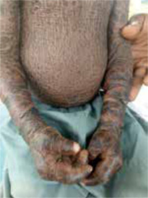
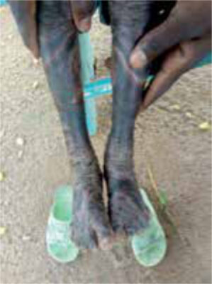
| Case A seven-year-old boy is presented by his parents for consultation. He has extremely dry skin that feels very firm and shows a scale-like structure over almost the whole body (see figure 1,2). The tight skin has formed an ectropion of both lower eyelids. He has had this from a young age, but it is unknown exactly for how long. One of his siblings has the same problem, but it is unknown if this is a brother or sister. His parents feel that he is not growing well. The child makes a sad impression. The parents are very worried about him and have been to different clinics/hospitals without a satisfactory result. |
Specialist advice
The dermatologists of the Consult Online panel diagnosed this as a form of ichthyosis, with a differential diagnosis of ichthyosis vulgaris and X-linked ichthyosis. The possible growth retardation could not be explained.
The specialists’ advice was to try and differentiate between the different forms of ichthyosis. This can be done by taking a broad (family) history to understand possible inheritance patterns or consanguinity, to exclude the condition as a possible syndromal sign (e.g. malignancy, see below), and understand the start and development of the disease from birth. Next to this, it is helpful to inspect the skin fully and describe the affected areas since this might also differentiate between different forms of ichthyosis. Making a definitive diagnosis makes it easier to predict inheritance and make a better assessment of the course of the disease.
General considerations for management
There is no cure for this condition, but symptoms can be treated. Treatment for both forms of ichthyosis is quite similar: taking baths with a bit of salt or bath oil on a regular basis. The skin needs to be kept oily by salving it multiple times a day with an emollient (oily ointment) or ureum on an oily base. Salicylic acid creams should not be used on the whole body, because of risk of systemic side-effects. Severe cases can be treated with systemic retinoids (acitretis or isotretinoin) but this is often not possible in a low-income setting. Additional extra-cutaneous manifestations should be treated. It is difficult to predict the effect of treatment but (some) improvement of skin and mucous membranes is possible over time. It is important to address the psychological impact of this disease. Counseling of possible pregnant carriers should be considered (see below).
Ichthyosis
An average adult has a skin surface of approximately 1.8 m2. The skin protects the body from influences of the external environment and is anatomically divided in three layers. The epidermis is the upper part and consists mainly of keratinocytes formed in the basal layer (stratum basale). The keratinocytes migrate upwards and undergo a complicated cornification process involving different enzymes and proteins before they form the outer (dead) part of the skin, the stratum corneum. Melanocytes, that form the melanin that pigments the skin, are also part of the epidermis. [1,2] The middle part is the dermis, consisting mainly of collagen, elastic tissue, vasculature (blood/lymphatic fluid), nerves for proprioception, pain and thermoregulation, hair follicles and glands. [1] The third layer called subcutis consists mostly of fatty tissue for isolation. [1]
Ichthyoses are a group of skin abnormalities, caused by cornification disorders, that lead to generalized scaling of the skin. [2,3] The severity of this scaling can vary among patients. [2] The majority of ichthyoses are inheritable; these can be divided in syndromal forms (affects skin and other organs) and non-syndromal forms (only cutaneous manifestations). The non-syndromal forms can be divided into common (ichthyosis vulgaris and X-linked ichthyosis) and uncommon forms (autosomal recessive congenital ichthyosis and other forms). [3] The heritable forms originate from mutations in different genes that encode for proteins and enzymes involved in skin development and function. [2] These mutations can lead to hyperplasia of the epidermis, thickening of the stratum corneum, increased desquamation and a scale-like structure of the skin, the result being a malfunctional skin barrier. [2,4]
Although most ichthyoses are inheritable, acquired forms do exist due to nutritional deficiencies, infections, autoimmune or malignant diseases. [2]
Ichthyosis vulgaris
Ichthyosis vulgaris is worldwide the most common form of ichthyosis, with a prevalence in the literature varying from 1:100 to 1:250 births. [2,3] Prevalence is worldwide but seems highest in Europe compared to Asia or even African Americans (lowest). [5]
The underlying cause is a loss-of-function mutation in the filaggrin-gene leading to abnormal cornification and trans epidermal loss of water. [3,4] Its inheritance pattern is autosomal ‘semi’ dominant, with incomplete penetrance. Individuals with the mutation on one allele (heterozygotes) show mild to moderate symptoms, whilst individuals with the mutation on both alleles (homozygotes) show moderate to severe symptoms. [2,3]
Symptoms start in the first months or years of life [3] and include dry skin with fine white-greyish scaling on the extensor surface of extremities and abdomen. The flexures and face are often not affected. In addition, there can be keratosis pilaris and hyperlinearity of the palms and soles. Symptoms tend to increase in dry and cold weather and decrease in hot, sunny and humid weather. It is strongly associated with atopy (eczema, asthma, and hay fever). [2,3]
| HERITABLE | |
|---|---|
| Syndromal forms (cutaneous + other organs) | Non-syndromal forms (only cutaneous) |
| Contiguous gene syndromes X linked dominant disorders With hair abnormalities With neurologic involvement With other associated symptoms | Common Ichthyosis vulgaris (1:250*) X-linked recessive ichthyosis** (1:6000*) Uncommon Autosomal recessive congenital ichthyosis (1:200,000*) lamellar ichthyosis congenital ichthyosiform erythroderma harlequin ichthyosis Keratopathic ichthyoses Other forms |
| ACQUIRED | |
| |
* incidence in number of births
** can present as syndromic and non-syndromic [7]
X-linked recessive ichthyosis
X-linked (recessive) ichthyosis is the second most common form of ichthyosis worldwide, with a prevalence in the literature varying from 1:4000 to 1:6000 male births. [2,3] Prevalence is worldwide and equally distributed among ethnic groups, almost exclusively in males.[6]
The condition is genetically transferred by an often asymptomatic female carrier that passes on a mutated X-chromosome to her male child, who will express symptoms of the disease. [3] The condition is caused by a mutation of the STS gene on the X-chromosome. This causes a steroid sulfatase deficiency and leads to abnormal cornification through different pathways. [2,3] In carrier females pregnant with a child with X-linked recessive ichthyosis, a steroid sulfatase deficiency in the foetal placenta leads to low or absent oestrogen levels in the intrauterine (urine and amniotic fluid) environment. This can cause insufficient cervical dilatation and decreased response to oxytocin. This may lead to serious obstetric complications such as prolonged or obstructed labour and the necessity of a caesarean section. [3,7]
Symptoms of X-linked recessive ichthyosis often start in the neonatal period with a systemic peeling of the skin and evolves within years into fine scaling and later on generalized, symmetrical, often dark brown adherent squama on the trunk, scalp, extremities, axillae, lateral parts of the face and especially the neck. Sparing can be seen in the popliteal and antecubital fossa, hand palms and foot soles and the central part of the face. [2,3,7] The affected area can itch. [7] This type of ichthyosis can also present as a syndromal form with extra cutaneous manifestations which include corneal opacities (that can also be seen in asymptomatic carriers), attention deficit hyperactivity disorder (ADHD), epilepsy and an increased risk of cryptorchidism. [2,3,7]
Differential diagnosis
Differentiating between these two forms of ichthyosis seems relevant to predict inheritance patterns, counsel possible carriers before or during pregnancy, and treat associated extracutaneous manifestations.
Often however, ichthyosis vulgaris and X-linked (recessive) ichthyosis are clinically indistinguishable because both forms show variations in symptoms. [3,7] As also advised by our specialists, it could be helpful to look at the pattern and colour of skin manifestations, obtain information about the onset and progression of the disease over time, including any problems during labour, ask for extracutaneous manifestations, and look for an inheritance pattern in the family history. Genetic testing could be performed for a more definitive diagnosis, this however is often impossible in a low-resource setting. [2,4]
Follow-up
The patient was prescribed an oily ointment (unscented Vaseline) to be used after soaking the skin in a bath. The patient was not seen again after this.
On review of the case while writing this report, his facial features and the large tongue are suggestive of Down’s syndrome. This condition is associated with ichthyosis vulgaris and may also explain the growth retardation.
Contact: c/o MTredactie@nvtg.org
References
Rodrigo-Nicolás B, Bueno-Martínez E, Martín-Santiago A, Cañueto J, Vicente A, Torrelo A, Noguera-Morel L, Duat-Rodríguez A, Jorge-Finnigan C, Palacios-Álvarez I, García-Hernández JL, Sebaratnam DF, González-Sarmiento R, Hernández-Martín A. Evidence of the high prevalence of neurological disorders in nonsyndromic X-linked recessive ichthyosis: a retrospective case series. Br J Dermatol. 2018 Oct;179(4):933-939.
Huidziekten. De Huid. [internet]. Available from: https://www.huidziekten.nl/folders/nederlands/huid.htm . [Accessed 11th October 2022].
Uptodate. Overview and classification of the inherited ichthyoses. [Internet]. Available from: https://www-1uptodate-1com-10019d2lx5e78.hanhosting.vakliteratuur.info/contents/overview-and-classification-of-the-inherited-ichthyoses?search=ichthyosis&source=search_result&selectedTitle=2~74&usage_type=default&display_rank=2 . [Accessed 11th October 2022].
van Vugt LJ, Steijlen PM, van Geel M, Vreeburg M, Gostyński A. ‘Common’ ichthyosis: belang van diagnostiek. Nederlands Tijdschrift voor Dermatologie en Venereologie. 2021 oktober;31(9):24-26.
Limmer AL, Nwannunu CE, Patel RR, Mui UN, Tyring SK. Management of Ichthyosis: A Brief Review. Skin Therapy Lett. 2020 Jan;25(1):5-7.
Uptodate. Ichthyosis vulgaris. [Internet]. Available from: https://www-1uptodate-1com-10019d28324d6.hanhosting.vakliteratuur.info/contents/ichthyosis-vulgaris?search=ichthyosis%20vulgaris&source=search_result&selectedTitle=1~15&usage_type=default&display_rank=1#H2986038674. [Accessed 17th October 2022].
Uptodate. Recessive X-linked ichthyosis. [Internet]. Available from: https://www-1uptodate-1com-10019d28324d6.hanhosting.vakliteratuur.info/contents/recessive-x-linked-ichthyosis?search=x%20linked%20ichthyosis&source=search_result&selectedTitle=1~15&usage_type=default&display_rank=1#H98140598. [Accessed 17th October 2022].
