Main content
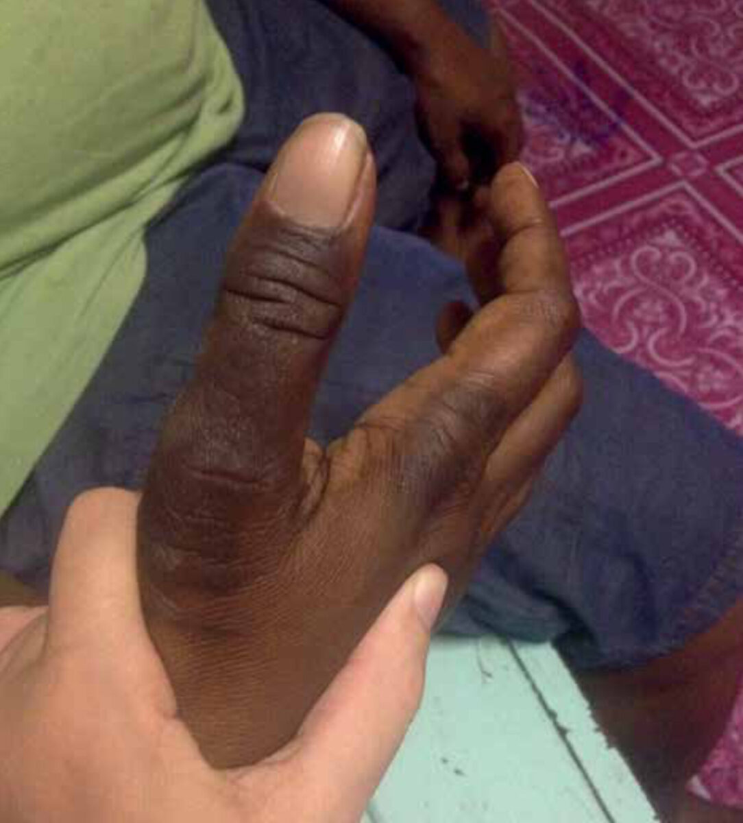
| Case A 35-year old woman without previous medical history presented with progressive intermittent stabbing pain in her right thumb since six years. The pain lasts for 10-30 seconds, several times a day, for a few days in a row. The pain is most intense just below the cuticle and radiates more proximally to the arm up to the shoulder. During an attack there seems to be loss of strength of the hand while sensitivity remains normal. Attacks are provoked by heavy work, sneezing and cold. Since three years, there is a dark discoloration of the skin on the dorsal side of the thumb and the skin is as thick as leather. There is a full range of motion in all joints. No other abnormalities are found during physical examination. |
Setting
This case is from Kikori District Hospital on the southwest peninsula of Papua New Guinea. The hospital has 80 beds and limited diagnostic capacity. Ultrasound is available, but there are no X-ray facilities and laboratory tests other than those used to diagnose HIV, syphilis, tuberculosis and malaria. Due to its location in the middle of the jungle, transport and therefore referral is difficult and only possible for those who can afford to travel.
Specialist advice
Neurologists and plastic surgeons were consulted for advice on this case, in particular on differential diagnosis and therapy.
The plastic surgeon mentioned, among other, a glomus tumour or osteoid osteoma as possible causes and suggested palpating the nail to look for discoloration and pain provocation, which is pathognomic for a glomus tumour. Furthermore, an X-ray of the thumb was advised to rule out osteoid osteoma although plain radiographs are not always diagnostic.
The discoloration of the skin could be due to the treatment of local healers or use of creams, the result of inflammation, or atrophy because of disuse of the thumb.
Follow-up
After putting pressure on the nail, a small blue discoloration was visible and recognizable pain was provoked, compatible with a glomus tumour. As treatment option of complete surgical excision was offered. However, as the patient was afraid to undergo surgery, she opted for a biopsy and to wait for the results. Two years later (!), the results of the biopsy, which was sent to Australia, came back. The pathology report stated: ‘Squamous hyperplasia in thick skin with some underlying and intrakeratotic haemorrhage. No fungi, parasites or neoplastic infiltrates are seen’. The intrakeratotic haemorrhage could result from the glomus tumour. The patient then agreed to surgical excision and is now waiting for a surgeon to visit the hospital.
Background of glomus tumour
A glomus tumour is a rare benign vascular neoplasm arising from a glomus body, a contractile vascular structure involved in thermoregulation in fingers, palms and soles that is highly concentrated in the subungual region. Hyperplasia of these vascular structures and smooth muscle cells can lead to swelling and cause pain provoked by pressure and histamine release in response to cold. In addition, non-myelinated nerve fibres penetrating the tumour are another potential source of pain. The cause of the hyperplasia is unknown, but subungual tumours have been associated with neurofibromatosis and reactive hyperplasia may be secondary to trauma. Middle-aged women are most at risk. [1]
Clinical features
A glomus tumour usually presents as a small bluish or pinkish-red, painful nodule or discoloration of the nail. The clinical triad of severe focal pain, pinpoint tenderness and cold hypersensitivity is highly suggestive. [2] Diagnosis is made clinically with help of three highly sensitive and useful tests. In Love’s pin test, pressure is applied on the tumour with a pinhead causing extreme pain. In Hildreth’s test, a tourniquet is applied along the arm to induce ischemia which will remove the pain. Another test is the cold-sensitivity test, by provoking pain when applying ice cubes or cold water on the tumour. [1]
Despite the classical presentation, delay in diagnosing these tumours for many years is common. Frequently patients are treated for neuropathic pain or hypersensitivity and undergo unsuitable treatments. The differential diagnosis of solitary painful tumour such as haemangioma, neuroma, leiomyoma or eccrine spiradenoma should be kept in mind. [1]
Imaging
A plain radiograph can show cortical thinning or erosive changes in the adjacent bone in some cases. Ultrasonography is useful in demonstrating the size, site and shape of the lesion but is highly influenced by the experience of the user and difficult in the subungual region. An MRI is most accurate in diagnosing patients with atypical symptoms and to prevent misdiagnosis. [1]
Histopathology
Histological features of glomus tumours include a variable composition of glomus cells, blood vessels, and smooth muscle cells. There are three types: solitary glomus tumours that are composed mainly of glomus cells, glomangioma characterized by the abundance of vessels, and glomangiomyoma with a predominance of smooth muscle cells. [2]
Therapy
Complete surgical excision is a must to achieve complete pain relief and to avoid recurrence. This can be approached from above after a nail extraction, but there is a high risk of permanent damage to the nail. Multiple modified surgical approaches have been described to prevent this complication. In the lateral approach, an incision is made close to the edge of the nail creating a large flap to reach the tumour avoiding excessive retraction of the nail. This flap is replaced after complete resection of the tumour. Although it is a benign tumour, it is necessary to remove it radically to avoid recurrence. In general, the prognosis is very good and most solitary glomus tumours do not recur. [1]
References
- Morey VM, Garg B, Kotwal PP. Glomus tumours of the hand: Review of literature. J Clin Orthop Trauma. 2016; 7:286-291.
- Sanchez IM, Ilkovitch D. A case of a glomus tumor presenting as an atypical hyperkeratotic papule of the hypothenar palm. JAAD Case Rep. 2017; 18:38-40. Onderkant formulier