Main content
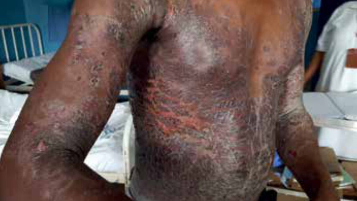
| Case A 23-year old man without previous medical history presented with rash throughout the body. It started three months ago with a rash on both hands which rapidly spread on arms, trunks and thighs. He started with Fluconazol which relieved the itch but not the rash. For the rash he visited multiple traditional healers who prescribed several different medications which he does not know the names of. At the moment of presentation, the patient had no itch but pain with movements which caused cracks in the skin. There was no fever. At physical examination, the skin was very dry with cracks with yellow exudates in the right flank. There were no further abnormalities found during physical examination. |
Setting
This case is from Biharamulo District Hospital, Kagera region, Tanzania. The hospital has 250 beds, and the medical staff consists of two medical officers and four assistant medical officers. They can carry out X-rays, ultrasounds and basic laboratory tests. The nearest referral hospital, in Mwanza, is 260 kilometres away.
Specialist advice
Dermatologists were consulted for advice on the differentials. They thought of toxic asteatotic eczema with infected cracks, probably caused by application of creams, and soap. They advised starting on antibiotics and topical therapy with strong paraffin or emulsifying ointment. Furthermore, they advised starting on prednisone 30 mg, assuming topical steroid ointment is very expensive in Tanzania and may not be available or affordable.
Follow-up
After five days of treatment and stopping with cleaning the skin with water, the skin looked much better and the pain was gone. After two weeks using prednisone, new itching skin lesions arose, consisting of annular lesions with an edge consisting of papules strung together. Again specialist advice was requested and the dermatologist thought of drug-induced lichen planus and advised taking a biopsy to differentiate between annular lichen planus, linear IgA dermatosis, and granuloma annulare. Unfortunately there was no possibility of taking a biopsy due to the costs and the patient was eventually started on a strong topical steroid ointment (clobetasol) combined with an antihistamine. After starting treatment, the patient did not show up for follow up. A cause of the lichen planus was therefore not identified, but it was known that he used diclofenac for pain relief and, due to the high incidence of malaria, it was conceivable that he had used antimalarial agents previously.

Background of lichen planus
Lichen planus is an inflammatory skin condition that affects 0.5-1% of the population in all age groups with no specific gender predominance. Lichen planus has a higher incidence in certain populations. In the U.S.: more African Americans were seen with lichen planus (72%) in comparison to the general clinic population (21%). In the UK, most children presenting with lichen planus originated from India (80.8%) in comparison to 28% of the city’s general child population. Furthermore, there is a predominance of lichen planus in children of Arab and Afro-Caribbean background.
The cause of lichen planus is unknown. Histologically, there is an autoimmune-mediated lysis and lymphocytic infiltrate in which cytotoxic T-lymphocytes act on the epidermis. There is an association with certain autoimmune diseases (thyroid diseases, rheumatoid arthritis, vitiligo, celiac disease, inflammatory bowel disease (IBD), primary biliary cirrhosis, alopecia areata and SLE), chronic liver diseases, particularly hepatitis C, and other viruses and bacteria, allergens and certain medications as discussed below.[1]
Clinical features
Classic lichen planus presents with the four Ps: purple, pruritus, polygon shaped and papules/plaques and are typically symmetric in distribution and affect any area on the body but rarely the face. There are many variants in morphology and location on the classic presentation which makes accurate diagnosis difficult. However, histopathological findings are largely consistent.[1]
Annular lichen planus
Approximately 3% to 7% of patients with lichen planus have the annular variant, with a predominance of male patients. It clinically presents as circular macules or plaques with raised borders with or without central atrophy with a diameter of 2-8 cm. Typically the lesions are localized but can be generalized.[1]
Drug induced lichen planus
Drug induced lichen planus is an uncommon cutaneous adverse effect. A wide range of medications have been associated with lichen planus and the list is growing as new agents are discovered and prescribed. Inducers include treatment with gold, antimalarial agents, ACE inhibitors, nonsteroidal anti-inflammatory agents, thiazide diuretics, penicillamine, dental amalgams, sulfasalazine, beta blockers, Viagra and proton-pump inhibitors. Compared to lesions of classic lichen planus, these are often larger in size, less monomorphic and more eczematous and associated with desquamation and crust. Typically it spares the classic sites of lichen planus such as the flexor surfaces, mucous membranes, nails and genitalia.[2] The time lag between the taking of the drug and the first cutaneous manifestations can be as long as three years, especially with penicillamine therapy, and depends on whether the patient has had previous exposure to the agent, dosages, host reaction and concurrent medication. Pruritus is frequent, although some patients are completely asymptomatic.[3]
Treatment
Treatment of drug induced lichen planus consists of removal of the offending agent. The eruptions resolve generally in a few weeks but can take as long as a year, and occasionally the eruptions recur intermittently despite discontinuation of the agent.[4] In case of prolonged disease, extensive disease or severe symptoms, treatment with topical or systemic corticosteroids may be warranted, although the efficacy has not been evaluated in randomized trials but is based on clinical experience and a few clinical studies.[5] After treatment the skin may become hyperpigmented skin which can take several years to disappear.[2]
References
- Weston G1, Payette M2. Update on lichen planus and its clinical variants. Int J Womens Dermatol. 2015 Sep 16;1 eCollection 2015 Aug.
- Jeremy Brauer MD, Henry J Votava MD, Shane Meehan MD, Nicholas A Soter MD. Lichenoid drug eruption. Dermatology Online Journal 15 (8):13
- Brauer J, Votava HJ, Meehan S, Soter NA. Lichenoid drug eruption. Dermatol Online J 2009; 15:13.
- Anderson TE. Lichen planus following quinidine therapy. Br J Dermatol 1967; 79:500.
- Le Cleach L, Chosidow O. Clinical practice. Lichen planus. N Engl J Med 2012; 366:723.