


Main content
Setting
This case originates from a general rural hospital in Kalongo, Uganda. The hospital has an inpatient capacity of 300 beds, distributed among 4 wards: Medical, Surgical, Maternity and Pediatrics. It has basic diagnostic facilities, including a laboratory, X-ray and ultrasound. The medical staff consists of 5 medical officers, a CEO and 2 specialists (1 surgeon, 1 doctor in tropical medicine). 30% of patients live below the poverty line; 40% live just above this threshold. The nearest referral hospital is a 2.5 hour drive by car.
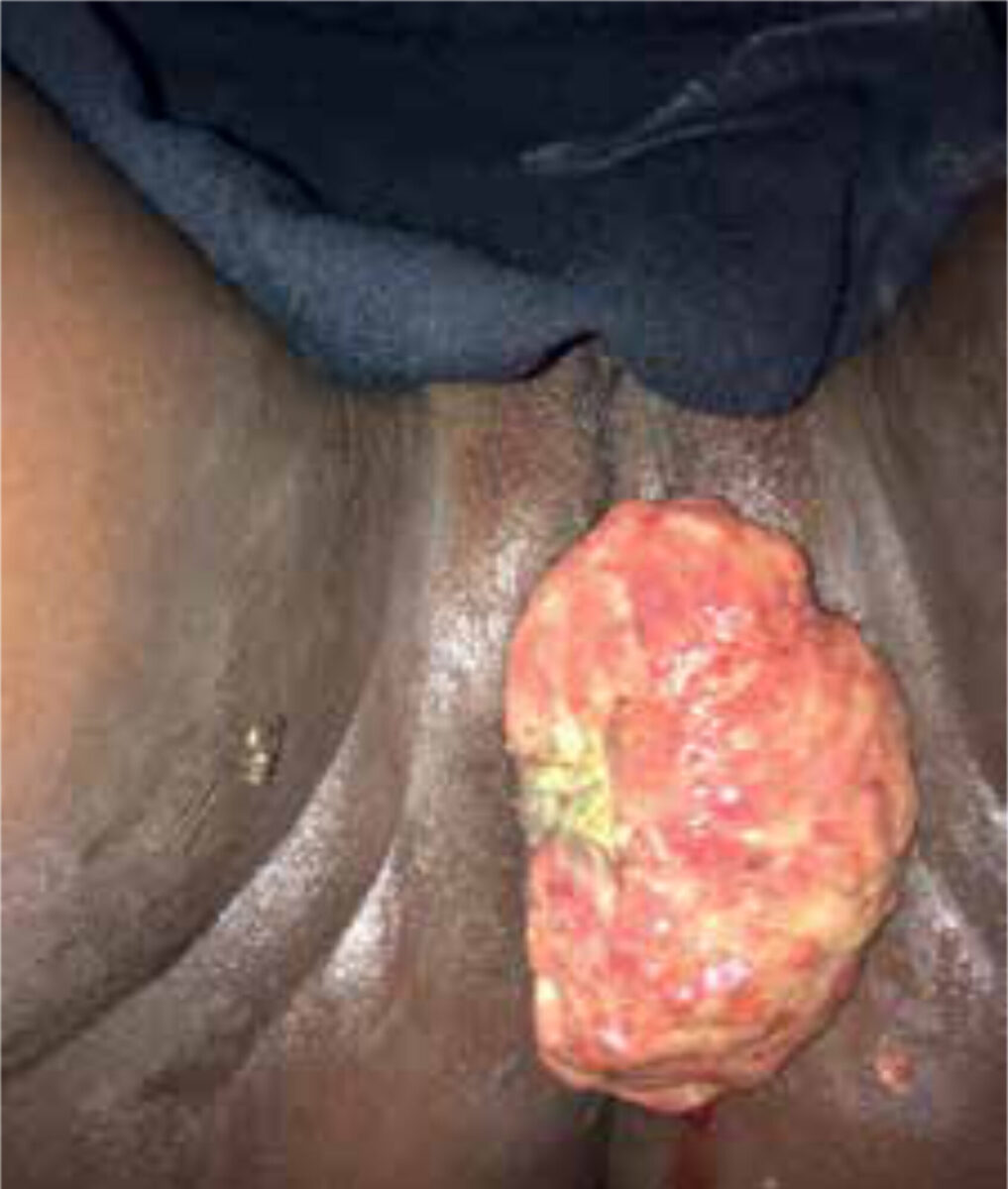
| Case A 15-year old girl presented at the outpatient department with a mass protruding from the vagina. The tumour had become apparent several days earlier, after experiencing the urge to defecate. The patient had a regular menstrual cycle. She did not appear ill during physical examination. On inspection of the genitalia, a malodorous, white-coated mass was seen. It was mobile and soft on palpation and did not seem adherent to the vulva. There was no vaginal discharge nor loss of blood. The mass itself was non-tender but examination was painful. Due to the pain and the fact that the girl was a virgin, vaginal internal examination was not performed. Therefore, the origin of the tumour was difficult to ascertain. An abdominal ultrasound was not helpful. It was decided to do further inspection and possible resection under anesthesia. |
Specialist advice
Gynaecologists were consulted for advice on the differentials. They suggested a few possible diagnoses, including a myoma nascens (however, both the young age and the fact that the tissue was soft on palpation was not typical), granulomatous tumour, venereal infection (however, the patient was not sexually active) or a caruncle originating from the urethra (which could be associated with Bilharzia).
Follow-up
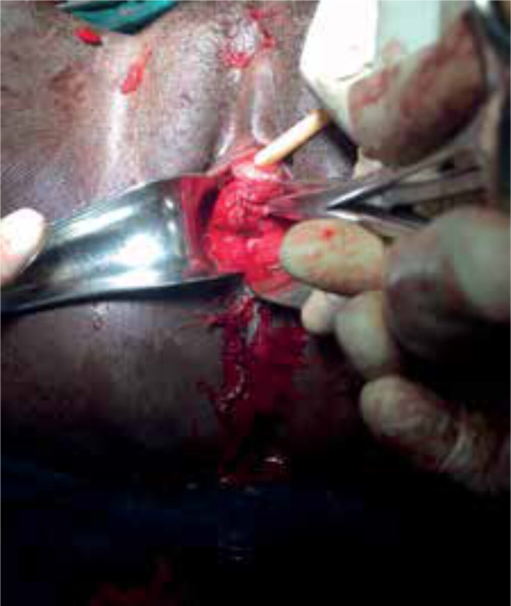
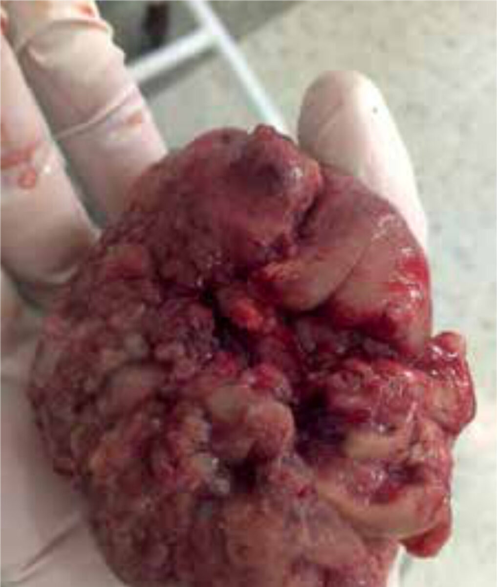
Inspection was performed under general anesthesia. Except for the mass protruding from the vagina, the vulva was normal on inspection. The meatus of the urethra was not affected. Vaginal walls were smooth and the mass was not adherent to the vagina. On palpation, the cervical os was identified, with the stalked tumour imbedded in the cervix. The tumour extended until about 3 cm proximal to the external os. It had two different consistencies, being soft on palpation at the front with central necrosis, but firm at the back. As resection in toto would entail resecting half the cervix, a partial resection of the mass was performed. The specimen was sent to Kampala for pathological analysis.
The pathology report stated: ‘Invasive tumour composed of ovoid cells with eosinophilic cytoplasm and atypical mitotic figures. The features are those of embryonal rhabdomyosarcoma.’
Several weeks after the partial resection, the patient came for follow-up and to discuss the further treatment plan. The primary treatment options for cervical rhabdomyosarcoma are radiotherapy and chemotherapy, both of which are only available in Kampala. However, at the time, radiotherapy was not possible as the machine was not working. Furthermore, the patient did not have the means to afford either radio- or chemotherapy. With the consent of the patient and her parents, a total abdominal hysterectomy was performed as best available treatment option in this setting. The uterus was sent to Kampala for pathologic examination.
The second pathology report stated the following: ‘Histological sections show a cervix partly infiltrated by spindle cell tumor. This is part of a rhabdomyosarcoma. The excision margins are free of tumor.’
The patient was advised to return for yearly follow-up, during a period of at least 10 years.
Background of rhabdomyosarcoma of the female genital tract
Pathophysiology and epidemiology
Rhabdomyosarcoma (RMS) is a malignant neoplasm. (¹) It accounts for 2-3.5% of cancers among children and adolescents, and it is the most common soft tissue sarcoma of early childhood. (1,3) RMS embryologically originates from skeletal muscle cells. It can occur anywhere in the body, but the most common sites are the head and neck region, the genitourinary tract and the extremities. (1,2) Tumours can be divided into four histologic subtypes: embryonal, pleomorphic, spindle cell and alveolar. (3) Of these, embryonal rhabdomyosarcoma ac-counts for 58% of all RMS and is therefore the most common. (1)
RMS of the genitourinary tract ac-counts for 3.5% of all RMS cases. (3) Of these, vaginal RMS usually occurs in the first decade of life, while cervical RMS commonly occurs in the second and third decades. Only 0.5% of RMS in girls originates from the cervix. (1) Presenting features are vaginal bleeding and/or a bulky mass.
Therapy and prognosis
RMS is most commonly treated by fertility sparing surgery, with neo-adjuvant chemotherapy and/or radiotherapy. Prognostic factors include the age of the patient, the histopathology of the tumour, the site of origin and the extent of the disease. (2) Following treatment, the overall 5 year survival rate for embryonal RMS is 73% and 47% in alveolar RMS. Vaginal lesions have a survival rate of 96%, as compared to 60% in cervical lesions. (2)
Take home messages
- RMS is a malignancy most commonly occurring in (early) childhood and adolescence.
- In RMS of the genitourinary tract, vaginal origin is most common and arises in the first decade of life.
- Cervical RMS is rare, mostly occurs in the second and third decades of life and has a worse prognosis.
- In high-resource settings, RMS can be treated with radiotherapy, chemotherapy and fertility sparing surgery.
References
- Narin MA, Karalok A, Basaran D, Turkmen O, Turan T, Tulunay G. Embryonal Rhabdomyo-sarcoma of the Cervix in Young Women. J Ado-lesc Young Adult Oncol. 2016;5(3):261-6.
- Pommert L, Bradley W. Pediatric Gynecologic Cancers. Curr Oncol Rep. 2017;19(7):44.
- Nasioudis D, Alevizakos M, Chapman-Davis E, Witkin SS, Holcomb K. Rhabdomyosarcoma of the lower female genital tract: an analysis of 144 cases. Arch Gynecol Obstet. 2017;20(6). [Epub ahead of print].
