
Main content
Nodular fasciitis is a benign soft tissue disease. Patients usually present with a rapidly growing soft tissue mass typically up to two centimetres in diameter. In this case report, an unusual presentation of nodular fasciitis will be discussed.
Case report
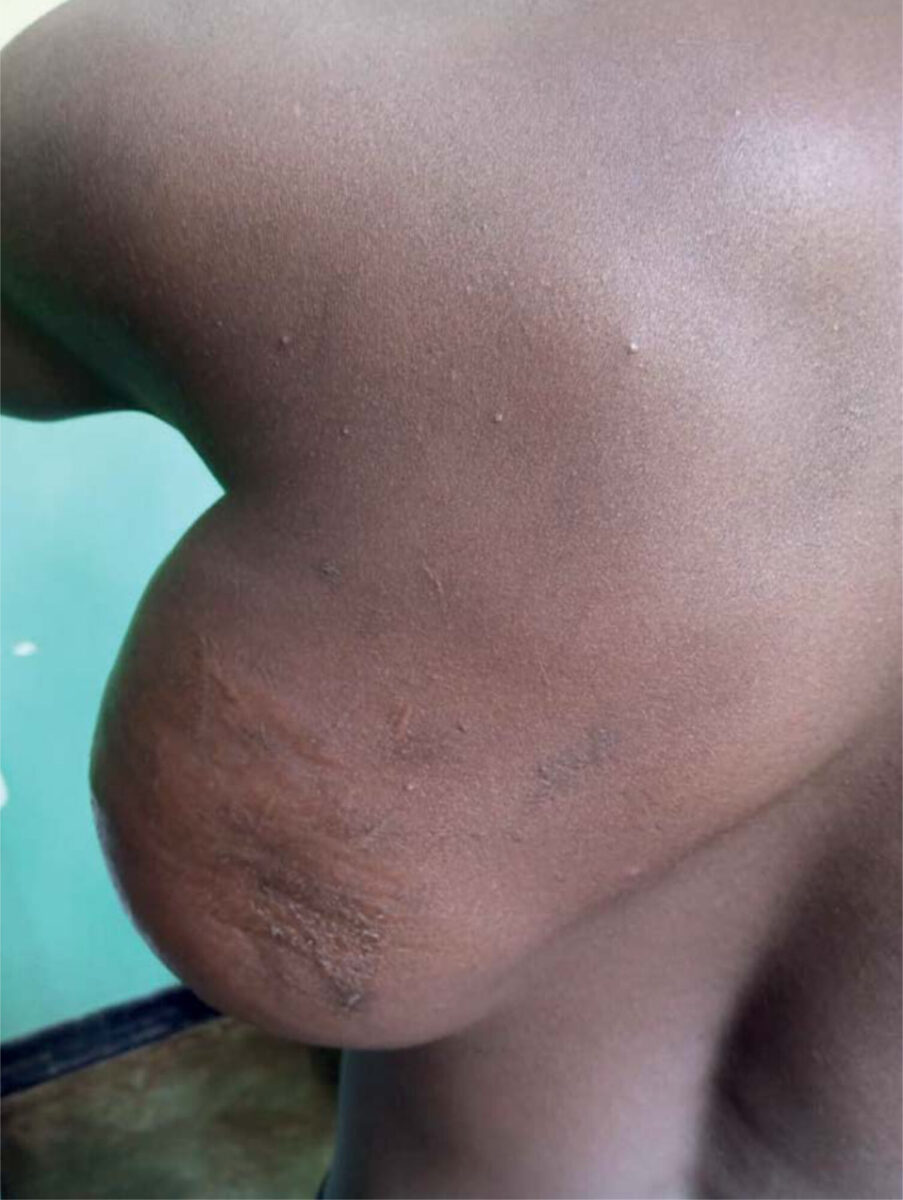
In May 2022, a 24-year-old, otherwise healthy man, presented to the Outpatient Department at Mulanje Mission Hospital, Malawi, with a mass in the left axillary region. The mass had been growing for the past two years. The size reduced the range of motion of the patient’s left upper extremity. The swelling was non-tender and there was no reported history of infection, trauma or other soft tissue masses. On examination, a firm and mobile soft tissue mass, measuring 15 by 15 cm was present on the left side of the trunk, just below the axilla. No skin lesions, erythema, calor or fluctuance were present. The differential diagnosis included a benign mass such as lipoma, fibroma, and epidermoid cyst. However, the size of the mass, the firm consistency, and the relatively rapid growth raised clinical suspicion of a malignant lesion, such as a sarcoma or neurofibroma. An ultrasound scan was performed and showed a firm mass, sonographically suggestive of a lipoma. A biopsy was taken and analysed at a private laboratory in Blantyre.
The findings upon histopathological examination were as follows. Sections showed proliferation of fibroblastic tissue forming storiform to solid patterns. The neoplastic cells showed a tissue culture pattern and extravasated red blood cells. No malignant cells were found. Findings were in line with the characteristics of nodular fasciitis.
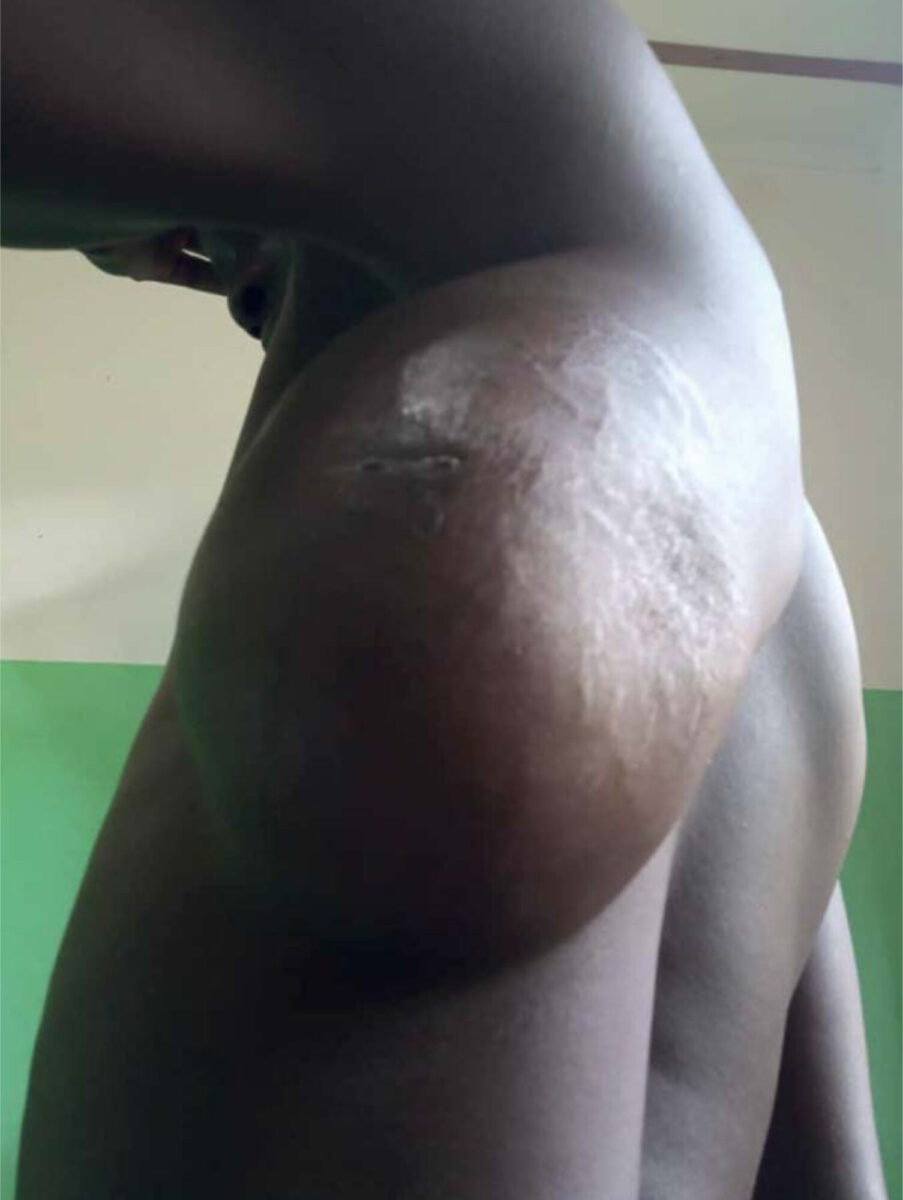
The histological findings were discussed with the tertiary surgical service in Blantyre. We were advised to excise and perform histopathological analysis of the mass. The mass was completely resected under general anaesthesia and a surgical drain was placed. 48 hours after resection, dark serous fluid was drained from the wound suspect for secondary wound infection. The incision was reopened, cloxacillin started, and the wound was dressed daily. After ten days, the infection had subsided. Following secondary skin closure, the patient was discharged home. Histopathological analysis of the resected mass was not performed due to financial constraints of the patient.
Upon a 3-month follow-up visit, the wound was found to have healed well without recurrence of a mass. The patient has not presented to the hospital since.
Discussion
Nodular fasciitis is a benign mesenchymal tumour. Other terms, such as infiltrative fasciitis, proliferative fasciitis, and pseudosarcomatous fasciitis, have been used synonymously.[1-4]
Clinical characteristics
Nodular fasciitis is most often observed in young adults (20 to 40 years of age).[1] Men and women are equally affected, but in childhood the mass is observed predominantly in boys. Most patients present with a rapidly growing, painless, solitary soft tissue mass. The consistency ranges from solid to nodular, rubbery or firm.[3] After a period of rapid growth, usually less than one month, the lesion tends to plateau in size and typically measures less than two to three centimetres.[4] However, masses with a size up to 10 to 15 centimetres exist as seen in this case. In atypical presentations, it is important to confirm the diagnosis of nodular fasciitis, as the clinical features share similarities with some forms of fibrosarcoma, which require a more aggressive treatment.[2] The most common site of presentation is the upper extremity, especially the forearm, followed by the chest wall, back, head and neck, with the latter presenting most often in infants and children.[3] The condition has also been reported on the hand, intra-oral, intra-neural, and intra-articular.[6-7]
When evaluating a mass like this, especially in remote settings, it is of great importance to differentiate between benign and malignant causes. Ideally, non-invasive diagnostic methods should be performed before any surgical resection is done. Imaging using MRI, CT, ultrasound, or PET-scan have all been used to describe the nature of such a lesion.[2-7] However, such diagnostic tools lack the capacity to definitely exclude malignancy and are often not available in small hospitals or those in low resource settings. Therefore, a biopsy is the recommended approach to obtain a preoperative diagnosis of a soft tissue mass when the nature is unclear. When a preoperative biopsy is performed, a postoperative histological analysis is of little added value. The preoperative approach is more cost-effective and this is of great importance in settings with limited resources.
The treatment of choice is surgical excision, although partial excision may be sufficient in nodular fasciitis as residuals may subsequently regress by scarring.[1, 2] The lesions do not metastasize and rarely reoccur. Reoccurrence of a lesion should lead to a critical review of the original diagnosis.[2, 3]
Conclusion
Nodular fasciitis is a rapidly growing, benign, soft tissue tumour which must be considered in the differential diagnosis of soft tissue masses. The preferred method of treatment is curative surgical excision with histopathological analysis to confirm the diagnosis.
References
- Enzinger FM, Weis SW. Soft tissue tumors. 2nd ed. St. Louis: CV Mosby; 1998. p. 102-112, 276-303.
- Bernstein KE, Lattes R. Nodular (pseudosarcomatous) fasciitis, nonrecurrent lesion: clinicopathologic study of 134 cases. Cancer. 1982 Apr 15;49(8):1668-78.
- Suh JH, Yoon JS, Park CB. Nodular fascitiis on chest wall in a teenager: a case report and review of the literature. J Thorac Dis. 2014 Jun;6(6) E108 -E110.
- Al-Hayder S, Warnecke M, Hesselfeldt-Niesen J. Nodular fasciitis of the face: a case report. Int J Surg Case Rep. 2019;61:207-209.
- Wang W, Huang Y, Wang C, Hong J, Ma C, Lin N, et al. Intra-articular nodular fasciitis: a rare lesion case report and an updated review of the literature. BMC Musculoskelet Disord. 2019 Jan 5;20(1):5.
- Domazet I, Njiric N, Jakovcevic A, Bitunjac A, Domazet K, Pašalić I, Mrak G. Intraneural nodular fasciitis of the dorsal scapular nerve: case report and review of the literature. J Neurol Surg A Cent Eur Neurosurg. 2021 Dec 12. 2023 Jul;84(4):404-407.
- Nair P, Barrett AW, Theodossy T. Oral nodular fasciitis: case report. Br J Oral Maxillofac Surg. 2004 Aug;42(4):360-2.