


Main content
Setting
This case is from Lolwa, a rural village in the Ituri rainforest located in the Democratic Republic of the Congo. A local Congolese medical officer and a medical doctor in global health and tropical medicine (MD GHTM) are running the hospital, which consists of 55 beds. The nearest option for advanced general surgery is a three-hour drive, but due to safety reasons patients are often not willing to travel.
| Case A healthy woman, gravida 3, para 1 (one spontaneous abortion) gave birth to a term neonate. She had three antenatal consultations during pregnancy, all of these without complications. She did not undergo any ultrasound examination during pregnancy. There was no evident history of abdominal trauma, although she reported occasional falls during her work on the fields. The family history consists of congenital clubfoot in the father’s family. During labour the membranes were artificially ruptured at 8 cm dilatation to improve the progression of labour. The amniotic fluid was red-brownish coloured and looked like old blood. There seemed to be more amniotic fluid than normal. After birth, the medical doctor was informed that the left arm was found amputated at the site of the proximal humerus, and the arm had been delivered after the head of the baby was born. At inspection, the amputated arm was oedematous and the hand looked smaller than the intact right hand of the neonate. At inspection of the neonate, the residual stump of the humerus was not completely covered with any tissue. There was also a bilateral clubfoot (Figures 1-4)*. The medical doctor in charge thought of an amniotic band syndrome and consulted the Consult Online panel for therapeutic advice on the residual stump and asked if antibiotic prophylaxis was indicated. |
Specialist advice
The diagnosis was confirmed by the specialists. They advised shortening the humerus until it was covered by soft tissue. But if the humerus does not or barely extends past the wound, healing per secundam (e.g. without surgical intervention) was suggested because of the lower risk of infection and to retain a maximum length of the stump. Antibiotic prophylaxis was recommended.
Amniotic band syndrome
Amniotic band syndrome or sequence (ABS) is a congenital disorder of foetal anomalies associated with foetal-placental fibrous bands. The aetiology is still unknown and considered to be multifactorial.[1,2] Different theories exist, suggesting either an intrinsic or an extrinsic cause. An intrinsic cause suggests that anomalies of the foetus result from the formation of amniotic bands due to a disturbance of the developing embryonic disk. An extrinsic cause suggests foetal entanglement occurs by mesodermal bands after disruption of the amnion.[2] The impact of ABS ranges from minor deformity to miscarriage or stillbirth. The incidence varies from 1:12,000 to 1:15,000 live births.[2]
The diagnosis of ABS can be made antenatally or, as in this case, peripartum. An antenatal diagnosis is suspected in the presence of amnion loose in the cavity, digital amputations, asymmetric limbs, or random anomalies during an ultrasound examination.[1,2] One should scan for amnion bands and see if movement of the foetus is restricted due to fixation. A foetal structural assessment is indicated in case of an amnion band. Clearly, all these findings are highly dependent on the quality of the ultrasound machine and the skills of the radiographer or radiologist. After antenatal diagnosis, timely intervention is recommended to release the amniotic bands via foetal surgery by fetoscopy to recover the perfusion of the affected area. Postnatal therapy consists of immediate surgical release of the bands or repair of the affected body part. Recurrence is rare, but some familial cases have been described. Club feet are associated with ABS, as is seen in this case.[3] To date, ABS is considered a sporadic event with no commonly accepted risk factors.
Follow-up
The neonate was admitted and the stump was covered with salinised compresses. The next day, the humerus was slightly shortened and covered with subcutaneous tissue and skin, using local anaesthesia and antibiotic prophylaxis. Wound healing was uncomplicated and shoulder movement intact. The correction of both club feet was done successfully using the Ponseti method.
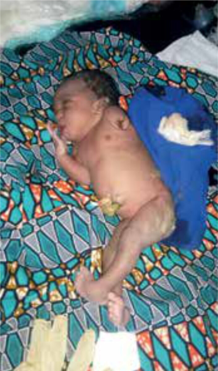
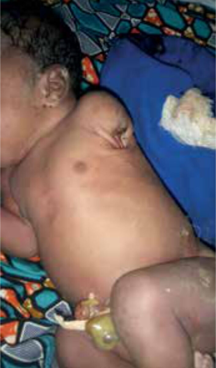
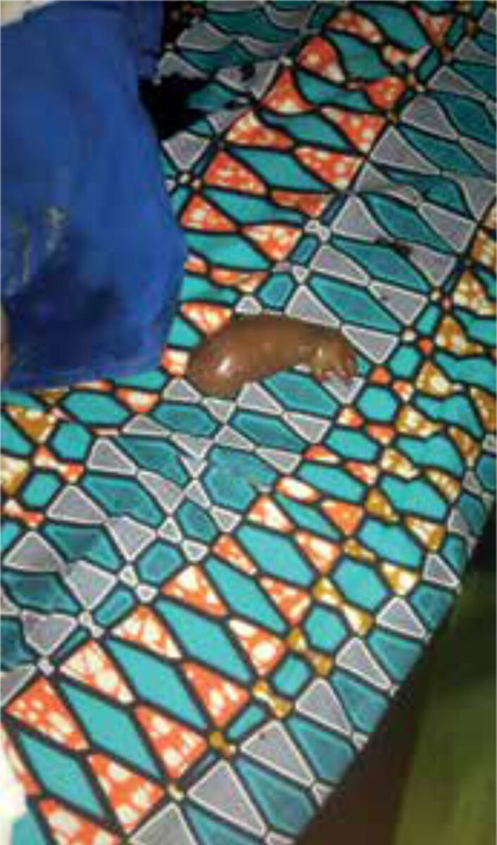
* Figures 1-4 published with the permission of the child’s mother.
References
- López-Muñoz E, Becerra-Solano LE. An update on amniotic bands sequence. Arch Argent Pediatr. 2018 Jun 1;116(3):e409-20. doi: 10.5546/aap.2018.eng.e409
- Gandhi M, Rac MWF, McKinney J. Amniotic Band Sequence. Am J Obstet Gynecol. 2019 Dec;221(6):B5-6. doi: 101016/j.ajog.2019.09.20.
- UpToDate® [Internet]. Alpen aan de Rijn: Wolters Kluwer; 2021. Bodamer OA, Amniotic band sequence; 2021 May 4. Available from: https://www.uptodate.com/contents/amniotic-band-sequence
